
Systems and methods to semi-automatically segment a 3D medical image using a real-time edge-aware brush

a technology of edge-aware brush and semi-automatic segmentation, applied in the field of improved learning systems and methods for medical image processing, can solve the problems of fewer staff, less skilled staff, and difficulty in effective management and use of imaging and information systems for examination, diagnosis, and treatment of patients, and achieve the effect of facilitating the generation of outputs

Active Publication Date: 2021-06-29
GENERAL ELECTRIC CO
View PDF27 Cites 2 Cited by
  • Summary
  • Abstract
  • Description
  • Claims
  • Application Information

AI Technical Summary

Benefits of technology

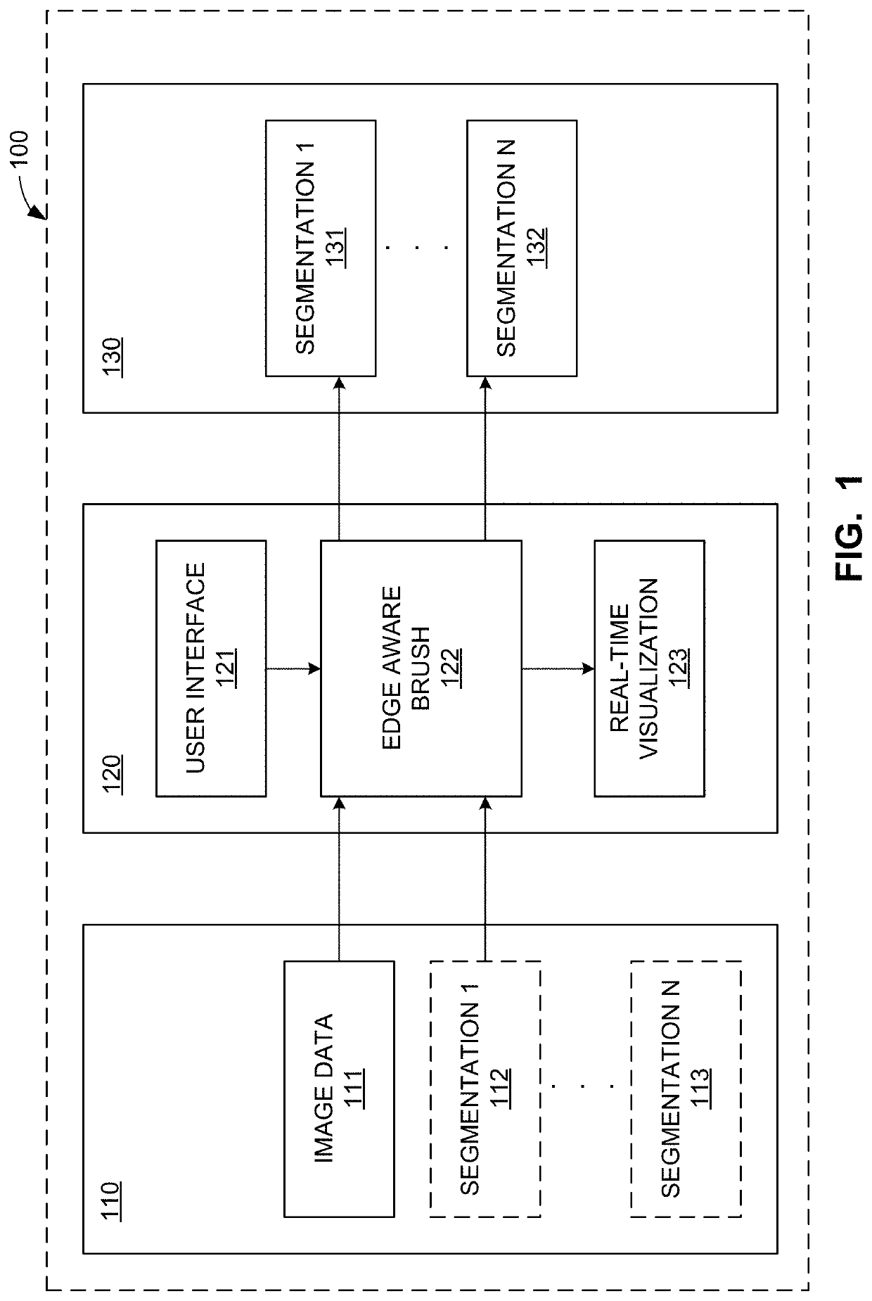
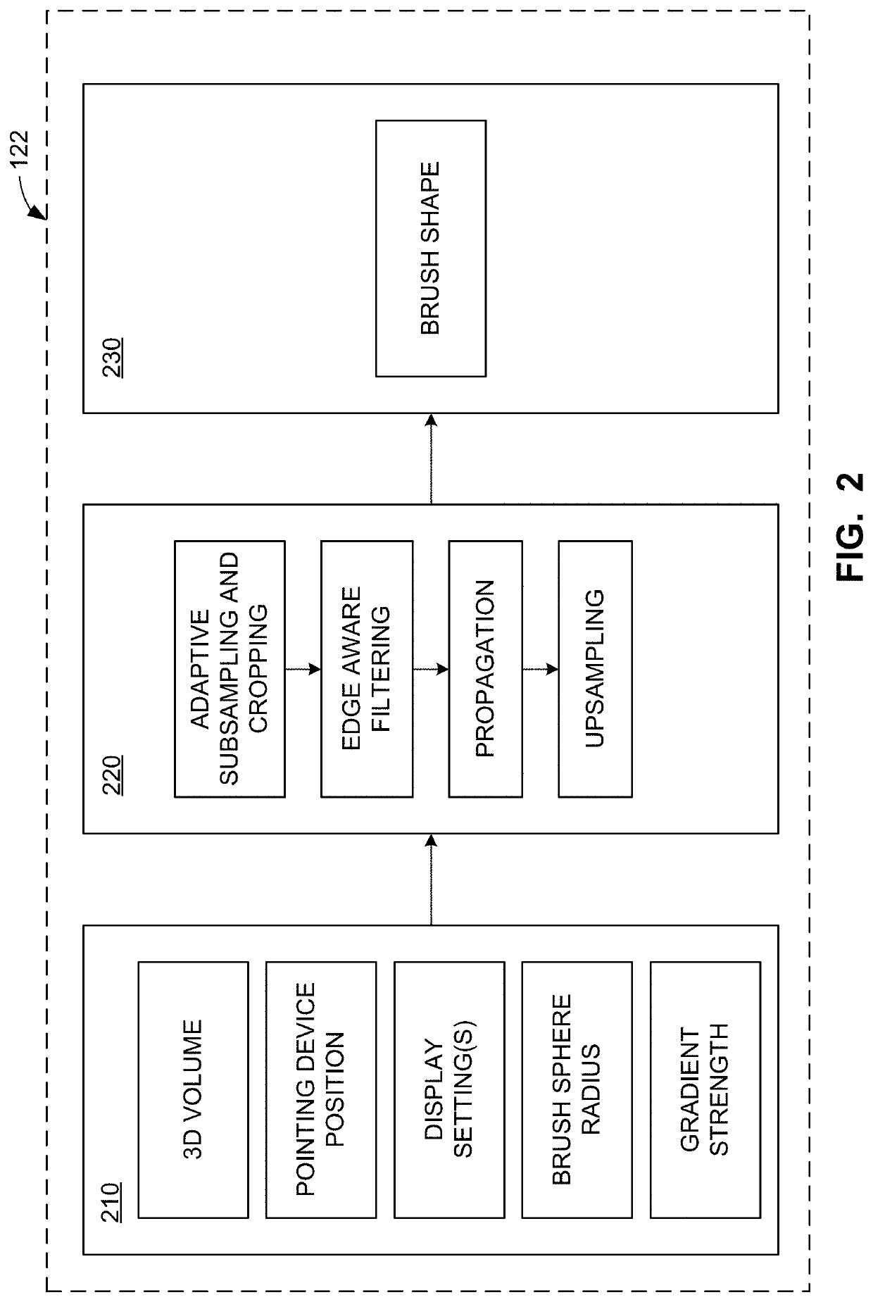
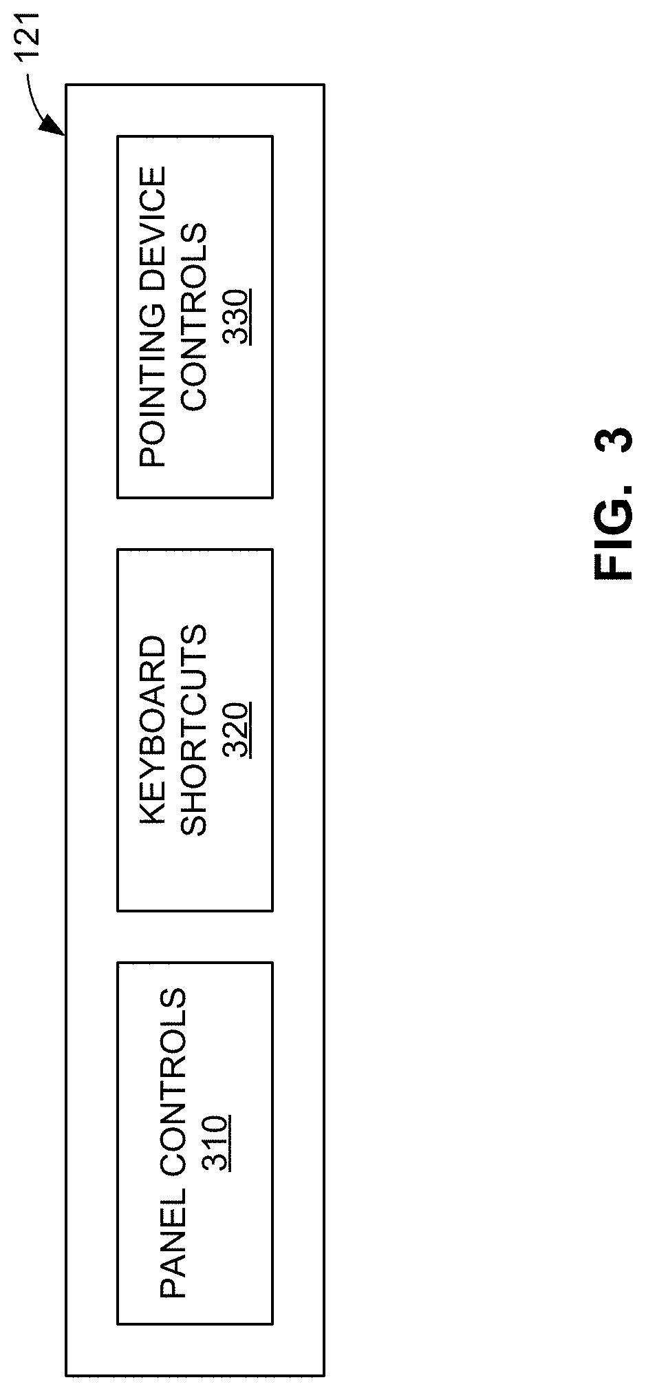
[0006]Certain examples provide at least one computer readable storage medium including instructions which, when executed, cause at least one processor to at least: construct a brush for segmentation of image data; provide an interactive representation of the brush with respect to the image data via a user interface, the interactive representation to be displayed and made available for interaction in each of a plurality of viewports provided for display of views of the image data in the user interface; enable update of the viewports based on manipulation of the representation; facilitate display of a preview of a segmentation of the image data corresponding to a location of the representation; and, when the segmentation is confirmed, facilitate generation of an output based on the segmentation.
[0007]Certain examples provide an apparatus including at least one processor and at least one computer readable storage medium including instructions. The instructions, when executed, cause the at least one processor to at least: construct a brush for segmentation of image data; provide an interactive representation of the brush with respect to the image data via a user interface, the interactive representation to be displayed and made available for interaction in each of a plurality of viewports provided for display of views of the image data in the user interface; enable update of the viewports based on manipulation of the representation; facilitate display of a preview of a segmentation of the image data corresponding to a location of the representation; and, when the segmentation is confirmed, facilitate generation of an output based on the segmentation.

Problems solved by technology

A variety of economy, operational, technological, and administrative hurdles challenge healthcare facilities, such as hospitals, clinics, doctors' offices, imaging centers, teleradiology, etc., to provide quality care to patients.
Economic drivers, less skilled staff, fewer staff, complicated equipment, and emerging accreditation for controlling and standardizing radiation exposure dose usage across a healthcare enterprise create difficulties for effective management and use of imaging and information systems for examination, diagnosis, and treatment of patients.
Healthcare provider consolidations create geographically distributed hospital networks in which physical contact with systems is too costly.
Healthcare provider (e.g., x-ray technologist, doctor, nurse, etc.) tasks including image processing and analysis, quality assurance / quality control, etc., are time consuming and resource intensive tasks impractical, if not impossible, for humans to accomplish alone.

Method used


Image

  • Systems and methods to semi-automatically segment a 3D medical image using a real-time edge-aware brush
  • Systems and methods to semi-automatically segment a 3D medical image using a real-time edge-aware brush
  • Systems and methods to semi-automatically segment a 3D medical image using a real-time edge-aware brush

Examples


Embodiment Construction

[0019]In the following detailed description, reference is made to the accompanying drawings that form a part hereof, and in which is shown by way of illustration specific examples that may be practiced. These examples are described in sufficient detail to enable one skilled in the art to practice the subject matter, and it is to be understood that other examples may be utilized and that logical, mechanical, electrical and other changes may be made without departing from the scope of the subject matter of this disclosure. The following detailed description is, therefore, provided to describe an exemplary implementation and not to be taken as limiting on the scope of the subject matter described in this disclosure. Certain features from different aspects of the following description may be combined to form yet new aspects of the subject matter discussed below.

[0020]When introducing elements of various embodiments of the present disclosure, the articles “a,”“an,”“the,” and “said” are i...


PUM


Abstract

Apparatus, systems, and methods to generate an edge aware brush for navigation and segmentation of images via a user interface are disclosed. An example processor is to at least: construct a brush for segmentation of image data; provide an interactive representation of the brush with respect to the image data via a user interface, the interactive representation to be displayed and made available for interaction in each of a plurality of viewports provided for display of views of the image data in the user interface; enable update of the viewports based on manipulation of the representation; facilitate display of a preview of a segmentation of the image data corresponding to a location of the representation; and, when the segmentation is confirmed, facilitate generation of an output based on the segmentation.

Description

FIELD OF THE DISCLOSURE[0001]This disclosure relates generally to improved medical systems and, more particularly, to improved learning systems and methods for medical image processing.BACKGROUND[0002]A variety of economy, operational, technological, and administrative hurdles challenge healthcare facilities, such as hospitals, clinics, doctors' offices, imaging centers, teleradiology, etc., to provide quality care to patients. Economic drivers, less skilled staff, fewer staff, complicated equipment, and emerging accreditation for controlling and standardizing radiation exposure dose usage across a healthcare enterprise create difficulties for effective management and use of imaging and information systems for examination, diagnosis, and treatment of patients.[0003]Healthcare provider consolidations create geographically distributed hospital networks in which physical contact with systems is too costly. At the same time, referring physicians want more direct access to supporting dat...

Claims


Application Information

Patent Timeline
Patent Type & Authority Patents(United States)
IPC IPC(8): G06K9/00G06T11/00H04N1/62G06T11/60G06T5/00G06F3/0484G06T5/20G06K9/40G09G5/02G06V10/25G06V10/44
CPCG06T11/001G06T5/003G06T5/005G06T11/60H04N1/62G06F3/04845G06K9/40G06T5/002G06T5/20G06T2207/20192G09G5/026G16H30/40G06T7/12H04N1/4092G06T7/155G06T2207/10072G06T2207/20092G06T2200/24G06F3/147G06V20/64G06V10/235G06V10/25G06V10/44G06V2201/03G06V10/30
Inventor MORARD, VINCENTGALLICO, TERRYGOGIN, NICOLAS
Owner GENERAL ELECTRIC CO
Learn More