X-ray imaging system

a technology of x-ray imaging and talbot imaging, which is applied in the direction of patient positioning for diagnostics, instruments, applications, etc., can solve the problems of difficult comparison of images, troublesome moving a subject between x-ray talbot imaging apparatus,

Active Publication Date: 2020-06-30
KONICA MINOLTA INC
View PDF8 Cites 1 Cited by
  • Summary
  • Abstract
  • Description
  • Claims
  • Application Information

AI Technical Summary

Problems solved by technology

However, it is troublesome to move a subject between an X-ray Talbot imaging apparatus and a normal X-ray imaging apparatus and to operate them individually.
Furthermore, since the subject is moved, the position of the subject in the field of view is different between an image obtained by the X-ray Talbot imaging and an image obtained by the normal X-ray imaging, and it is difficult to compare the images.

Method used


Image

  • X-ray imaging system
  • X-ray imaging system
  • X-ray imaging system

Examples


Embodiment Construction

[0027]Hereinafter, one or more embodiment of the present invention will be described with reference to the drawings. However, the scope of the invention is not limited to the disclosed embodiments.

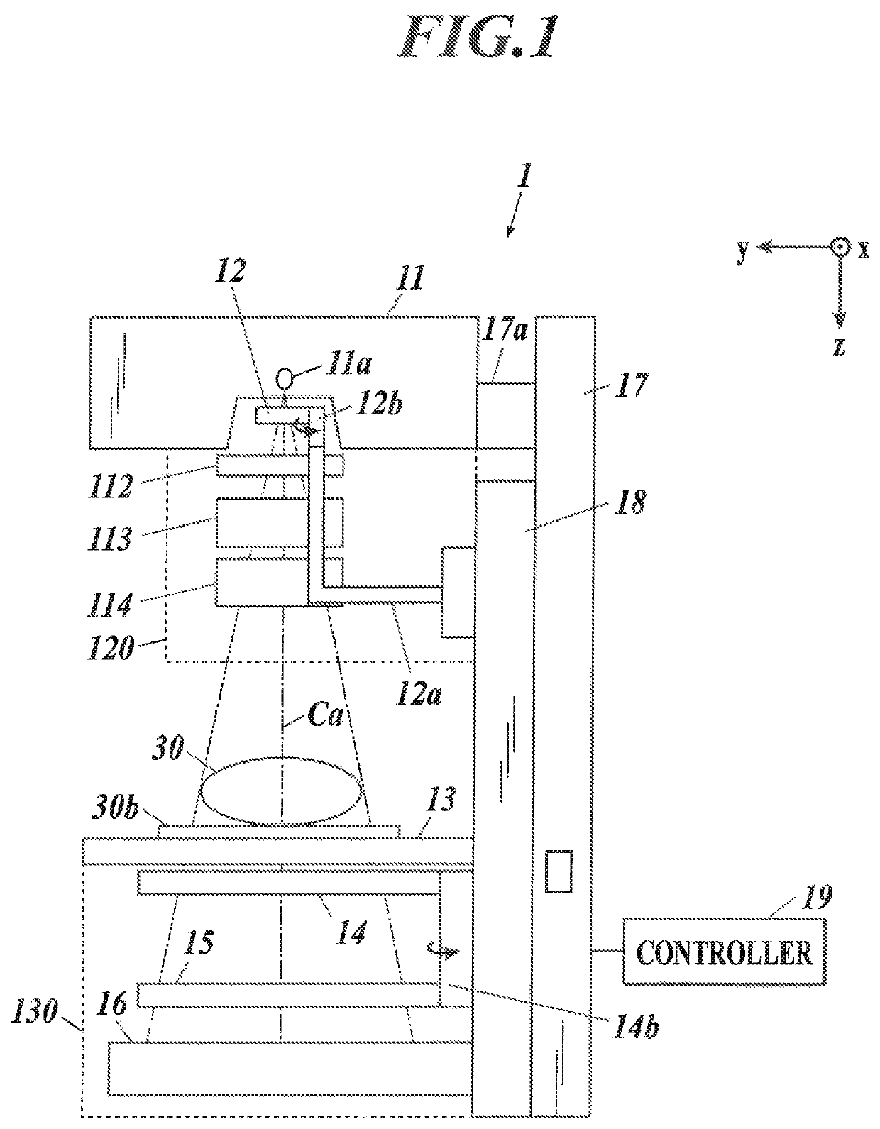
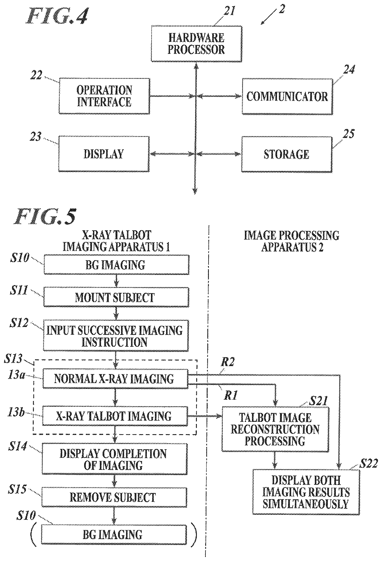
[0028]The embodiment illustrates an X-ray imaging system in which an X-ray Talbot imaging apparatus 1 radiographs a subject 30, and an image processing apparatus 2 generates a plurality of reconstruction images, including at least three reconstruction images of an absorption image, a differential phase image and a small-angle scattering image, from a moire image taken by the X-ray Talbot imaging apparatus 1. Further, the X-ray imaging system is also used to obtain a normal X-ray radiographic image. Examples of the subject 30 include human bodies (mainly for medical purposes), animals and plants, and goods to be subjected to non-destructive testing.

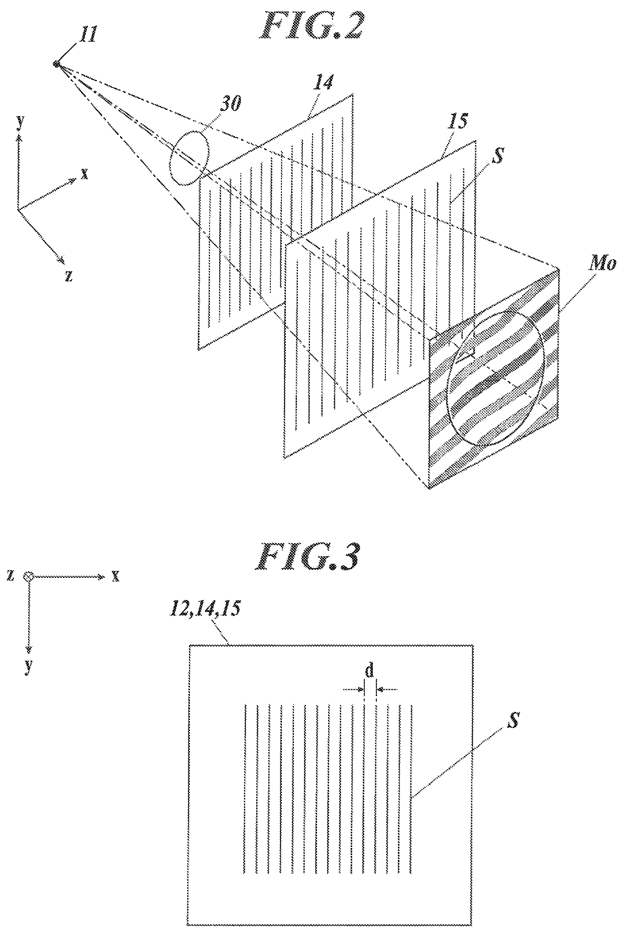
[0029]As the X-ray Talbot imaging apparatus 1 in this embodiment, one that includes a Talbot-Lau interferometer provided with a source grating (...


PUM

PropertyMeasurementUnit
pixel sizeaaaaaaaaaa
thicknessaaaaaaaaaa

Abstract

An X-ray imaging system includes an X-ray Talbot imaging apparatus and an image processing apparatus. The image processing apparatus generates reconstruction images from the moire image taken by the X-ray Talbot imaging apparatus. The reconstruction images include a differential phase image, an absorption image and a small-angle scattering image. The X-ray imaging system performs X-ray Talbot imaging and normal X-ray imaging. X-ray Talbot imaging includes setting an exposure energy of the X-ray Talbot imaging apparatus to a first exposure energy and taking the moire image with the X-ray detector. Normal X-ray imaging includes setting the exposure energy of the X-ray Talbot imaging apparatus to a second exposure energy and taking an absorption contrast image with the X-ray detector by normal X-ray imaging.

Description

BACKGROUND1. Technological Field[0001]The present invention relates to an X-ray imaging system.2. Description of the Related Art[0002]Almost all X-ray images for medical purposes to make a diagnosis and for non-destructive testing to inspect the inside of an object are based on an absorption contrast technique. The absorption contrast technique is to create contrast according to difference in attenuation of X-ray that passes through a subject. Meanwhile, a phase contrast technique is also used which is to create contrast according to phase change of X-ray instead of absorption of X-ray.[0003]One of phase contrast imaging techniques is a Talbot imaging technique that utilizes the Talbot effect. As an example of the Talbot imaging technique, an X-ray imaging apparatus equipped with a Talbot interferometer or a Talbot-Lau interferometer with a one-dimensional grating (hereinafter referred to as an X-ray Talbot imaging apparatus) has been known in the art. By subjecting a moire image ta...

Claims


Application Information

Patent Timeline
Patent Type & Authority Patents(United States)
IPC IPC(8): A61B6/00A61B6/04G01N23/041A61B6/12G01N23/04
CPCA61B6/4291A61B6/4417G01N23/041A61B6/4035A61B6/545A61B6/04A61B6/5205A61B6/5235A61B6/482A61B6/463A61B6/12A61B6/484A61B6/502G01N2223/401G01N23/04
Inventor KITAMURA, MITSUHARU
Owner KONICA MINOLTA INC
Why Eureka
Try Eureka