Eureka AIR delivers breakthrough ideas for toughest innovation challenges, trusted by R&D personnel around the world.

Fast dual energy for general radiography

a dual energy and general radiography technology, applied in the field of general radiography, can solve the problems of blurring and/or artifacts in the final image, delay between the two images, etc., and achieve the effect of improving and accurate capturing of image information

Active Publication Date: 2018-11-27
GENERAL ELECTRIC CO
View PDF34 Cites 0 Cited by
  • Summary
  • Abstract
  • Description
  • Claims
  • Application Information

AI Technical Summary

Benefits of technology

[0007]A technical effect of some embodiments described herein may be an improved and accurate capturing of image information. Embodiments may be associated with systems and / or computer-readable medium storing instructions to perform any of the methods described herein.

Problems solved by technology

Note, however, that two images need to be generated (low energy and high energy), and there may be a delay between the two images due to the X-ray tube switching and detector read out time (e.g., a 150 ms delay).
This could cause mis-registration between the two images because of patient / anatomy motion in that time period, which can cause blurring and / or artifacts in the final image.

Method used


Image

  • Fast dual energy for general radiography
  • Fast dual energy for general radiography
  • Fast dual energy for general radiography

Examples


Embodiment Construction

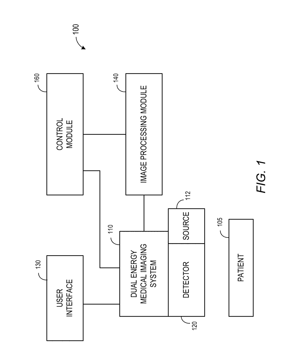
[0021]FIG. 1 illustrates a system 100 using dual energy image acquisition in accordance with some embodiments. The system 100 comprises a patient 105 and a dual energy medical imaging system 110. The dual energy medical imaging system 110 comprises a detector 120, a user interface 130, an X-ray source 112, an image processing module 140, and a control module 160. In some embodiments, the dual energy medical imaging system 110 may be adjusted quickly for changes in imaging techniques.

[0022]The detector 120 converts X-rays to digital signals. The detector 120 may be, for example, a solid state detector. The detector 120 may adjust its operation relatively quickly (e.g., within 1 second). The dual energy medical imaging system 110 employs the detector 120 to produce an image based on energy, such as X-ray energy, transmitted through the patient 105.

[0023]The user interface 130 allows a user to input configuration information. The configuration information may include information such a...


PUM

PropertyMeasurementUnit
voltageaaaaaaaaaa
energyaaaaaaaaaa
voltageaaaaaaaaaa

Abstract

Some embodiments are associated with an X-ray source configured to generate X-rays directed toward an object, wherein the X-ray source is to: (i) generate a first energy X-ray pulse, (ii) switch to generate a second energy X-ray pulse, and (iii) switch back to generate another first energy X-ray pulse. A detector may be associated with multiple image pixels, and the detector includes, for each pixel: an X-ray sensitive element to receive X-rays; a first storage element and associated switch to capture information associated with the first energy X-ray pulses; and a second storage element and associated switch to capture information associated with the second energy X-ray pulse. A controller may synchronize the X-ray source and detector.

Description

BACKGROUND[0001]The invention relates to general radiography and, more particularly, to methods and systems providing fast Dual Energy (“DE”) radiography for medical imaging and similar applications.[0002]Generally, an X-ray generated by an X-ray tube may pass through a patient and then be detected by an x-ray detector. Detected X-ray levels may then be used to generate an image of the patient. To improve the quality of the image, two exposures of X-rays may be utilized, each at a different energy level. Such an approach is referred to as “dual energy” radiography.[0003]One objective of dual energy radiography is to obtain images that enhance contrast separation within the image by utilizing two scans at these different energy spectra. For example, a dual energy approach may facilitate an extraction of bone information or soft tissue information—or separate any two different arbitrary materials. There is also evidence that dual energy can improve coronary artery calcium detection, w...

Claims


Application Information

Patent Timeline
Patent Type & Authority Patents(United States)
IPC IPC(8): A61B6/00G01N23/087H01L27/146
CPCA61B6/4233A61B6/482A61B6/405A61B6/4208H01L27/14658A61B6/542G01N23/087H01L27/14641A61B6/40A61B6/42A61B6/50A61B6/504A61B6/505A61B6/5264G01N2223/423G01N23/044
Inventor ZOU, YUNSABOL, JOHN MICHAELYANOFF, BRIAN DAVIDLAI, HAOJACOB, BIJUNYE, KATELYN ROSECHEN, FENG
Owner GENERAL ELECTRIC CO
Why Eureka
Eureka Blog
Learn More