Visual rectal tumor insection edge positioning device

A positioning device and technology for rectal tumors, applied in the field of medical devices, can solve the problems of inaccuracy, reduce the recurrence probability of anastomotic tumors, blur the distance between the distal margin and the tumor, and achieve better and clearer observation.

Inactive Publication Date: 2022-08-05
PEOPLES HOSPITAL PEKING UNIV +1
View PDF6 Cites 0 Cited by
  • Summary
  • Abstract
  • Description
  • Claims
  • Application Information

AI Technical Summary

Problems solved by technology

[0002] In the surgical operation of malignant tumors of the digestive tract, it is often necessary to ensure that the upper and lower resection margins of the tumor are far enough away from the tumor to ensure that the pathological examination of the resection margin is negative, so as to greatly reduce the probability of tumor recurrence at the anastomotic stoma. 》, for different stages of rectal cancer, the distance between the distal resection margin and the tumor has been clarified, but there is still no surgical instrument that can help doctors measure the above distance during surgery, especially laparoscopic surgery, which is extremely limited, only The above-mentioned distance can be roughly judged by relying on some laparoscopic surgical instruments as reference objects. So far, the distance between the distal resection margin and the tumor in laparoscopic surgery is vague and inaccurate

Method used


Image

  • Visual rectal tumor insection edge positioning device
  • Visual rectal tumor insection edge positioning device
  • Visual rectal tumor insection edge positioning device

Examples


Embodiment 1

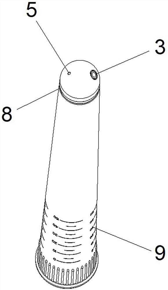
[0028] The marking mechanism is a light-emitting portion disposed on the peripheral side of the front end of the inner core 2 , and the light-emitting portion is flush with the zero-scale line of the front scale area 4 in an initial state.

[0029] The light-emitting part is an annular light strip 8 .

[0030] The scale area includes a rear scale area 9 disposed on the outer wall of the rear side of the inner core 2 , and the rear scale area 9 is used to display the relative distance between the light-emitting portion and the start portion of the guide tube 1 .

[0031] The rear scale area 9 is indicated by the rear end of the guide tube 1 . The relative distance between the light-emitting portion and the starting portion can be directly determined by observing the scale of the rear scale area 9, and then the distance between the light-emitting portion and the lower tumor margin can be determined.

[0032] The working principle of embodiment 1 is as follows:

[0033] 1. Inse...

Embodiment 2

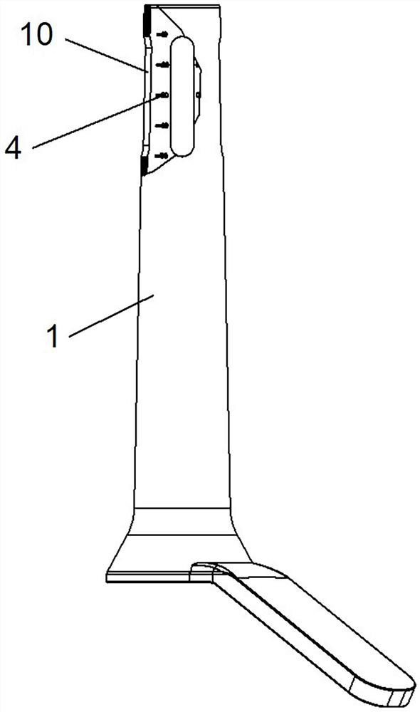
[0038] The marking mechanism is a window 10 opened on the peripheral wall of the guide tube 1 .

[0039] The window 10 is a strip-shaped through hole, and the strip-shaped through hole extends along the length direction of the front scale area 4 .

[0040] A plurality of the windows 10 are arranged in total, and each window 10 is symmetrically distributed in a ring around the central axis of the guide tube 1 .

[0041] The window 10 is located at the 1-5 cm scale line of the front scale area 4 of the guide tube 1 .

[0042] In this embodiment, a total of four windows 10 are provided, which are respectively located at the 12 o'clock, 3 o'clock, 6 o'clock and 9 o'clock positions of the guide tube.

[0043] The working principle of embodiment 2 is as follows:

[0044] 1. Insert the visual rectal tumor lower margin positioning device into the rectum through the anus to reach the lower margin of the tumor.

[0045] 2. After confirming that the starting part of the front end of t...


PUM


Abstract

The invention relates to a visual rectal tumor undercut edge positioning device. The guide pipe is used for positioning the tumor insection edge, and the inner core penetrates through the inner side of the guide pipe in a sliding mode. The front end of the inner core is provided with a camera unit, the front end of the guide tube is provided with a starting part used for abutting against the insection edge of a tumor, a scale area is arranged between the guide tube and the inner core and used for displaying the linear sliding distance of the inner core relative to the starting part, and the guide tube or the inner core is provided with a marking mechanism used for assisting in marking the rectum. The method has the advantages that a doctor can clearly quantify the excision range in an operation, difference caused by personal judgment is avoided, and the situation of excessive excision or insufficient excision is avoided. Specifically, an operator can directly judge the position of the undercut edge in the rectum cavity through scales and calibrate the position of the undercut edge on the inner surface or the outer surface of the rectum cavity.

Description

technical field [0001] The invention relates to the field of medical devices, in particular to a visual rectal tumor lower incision margin positioning device. Background technique [0002] In the surgical operation of malignant tumors of the digestive tract, it is often necessary to ensure that the upper and lower resection margins of the tumor are far enough away from the tumor to ensure that the pathological examination of the margins is negative, thereby greatly reducing the recurrence probability of anastomotic tumors. ", for different stages of rectal cancer, the distance between the distal resection margin of the tumor and the tumor is clarified, but there is still no surgical instrument that can help doctors measure the above distance during surgery, especially laparoscopic surgery, which is extremely limited, only The above distance can be roughly judged by relying on some laparoscopic surgical instruments as reference objects, so far, the distance between the distal...

Claims


Application Information

Patent Timeline
Patent Type & Authority Applications(China)
IPC IPC(8): A61B1/31A61B1/04A61B17/02A61B17/94
CPCA61B1/31A61B1/04A61B17/02A61B17/00234A61B1/00071
Inventor 曹键叶颖江莫易凡林聪杰邱木旺林龙杰苏占明
Owner PEOPLES HOSPITAL PEKING UNIV
Why Eureka
Try Eureka