
Body composition automatic measurement system based on abdomen CT image and deep learning

A CT image, automatic measurement technology, applied in image analysis, image data processing, medical automatic diagnosis, etc., can solve the problems of small number of samples, small sample size, inaccurate segmentation results, etc.

Pending Publication Date: 2022-04-12
FUDAN UNIV
View PDF0 Cites 0 Cited by
  • Summary
  • Abstract
  • Description
  • Claims
  • Application Information

AI Technical Summary

Problems solved by technology

The disadvantages of these two methods are: such methods are difficult to distinguish tissues under certain special pathological conditions, resulting in inaccurate segmentation results; no matter whether all skeletal muscles are analyzed as a whole or only psoas muscles are analyzed, CT is not fully utilized. Imaging; more importantly, using only skeletal muscle on L3 or L4 single-axial CT slices, the sample size is small and muscle mass cannot be accurately described
This measurement lacks an association analysis of different types of skeletal muscle with sarcopenia and uses only skeletal muscle on a single axial CT slice, with a small sample size

Method used


Image

  • Body composition automatic measurement system based on abdomen CT image and deep learning
  • Body composition automatic measurement system based on abdomen CT image and deep learning
  • Body composition automatic measurement system based on abdomen CT image and deep learning

Examples


Embodiment Construction

[0061] The present invention will be further described below in conjunction with examples and system overall framework drawings.

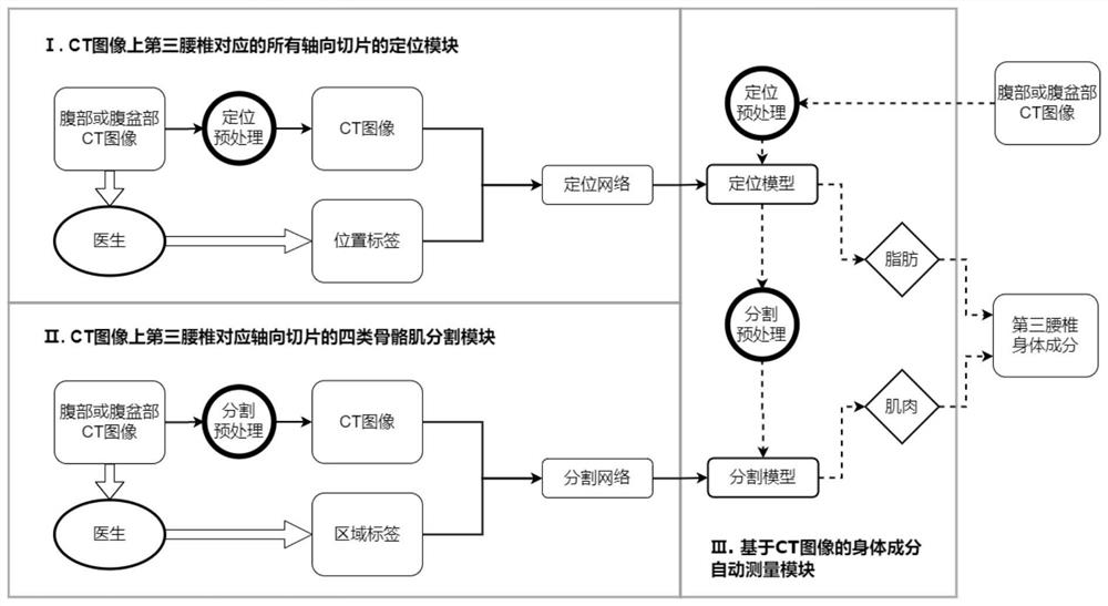
[0062] Take the clinical original image from clinical collection as an example, the present invention automatically measures the body component application process, such as Figure 8 Indicated.

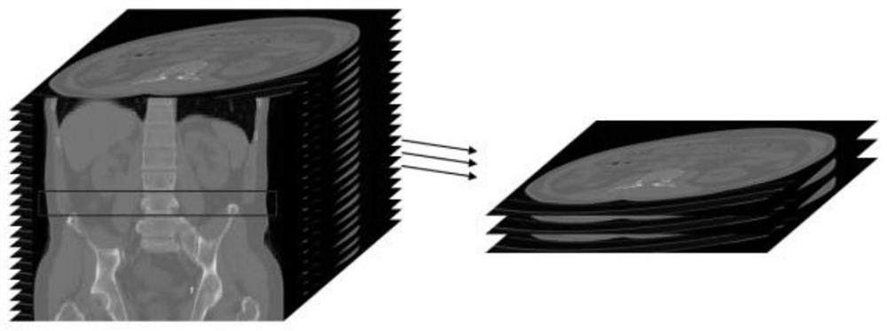
[0063] Module 1 screens the axial slice corresponding to the third lumbar vertebrae. First, 101 clinical CT image sets of 101 non-hepatic hardening patients are positioned, so that the three-dimensional data set is stored as an extension called "PNG format", and random vertical flips and affine transformations are performed. All slices were input to the positioning model obtained by 216 cases of liver hardening data sets to obtain the predicted results of the third lumbar slit, as shown in Table 1. "0" in Table 1 shows an axial slice corresponding to the third lumbar vertebrae, "1" indicates the axial slice corresponding to the third lumbar vertebrae. Accuracy ...


PUM


Abstract

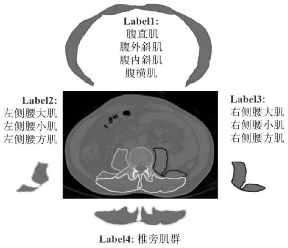
The invention discloses a body composition automatic measurement system based on an abdominal CT image and deep learning. The system comprises a positioning module of all axial slices corresponding to the third lumbar vertebra on the CT image, a four-class skeletal muscle segmentation module of the axial slices corresponding to the third lumbar vertebra on the CT image, and a body composition automatic measurement module based on the CT image. The first two modules are used for training a deep learning model to position and segment four types of skeletal muscles of the third lumbar vertebra, and the proportion of body components such as fat and muscles is automatically calculated. According to the skeletal muscle segmentation method based on CT data of clinical cirrhosis patients, the average Dice of the four types of skeletal muscle segmentation results reaches 0.9283, and the average surface distance is 0.6779 mm. Body components such as subcutaneous fat and intra-abdominal fat can be obtained through threshold treatment. According to the method, the time for obtaining the body components corresponding to the third lumbar vertebra in batches is 2-3 seconds, and the method has important significance on clinical diagnosis of complications of liver cirrhosis patients.

Description

Technical field [0001] The present invention belongs to the technical field of medical diagnostic equipment, and is specifically related to the body component automatic measurement system based on the abdominal CT image. Background technique [0002] Body ingredient analysis is to quantify muscle and adipose tissue. Its measurements are typically based on biological parameters or medical images. In particular, the skeletal muscle mass on the axial CT slice of the Third Lumbar Vertebra, L3 is measured, proves to be the most accurate [1]. The skeletal muscle is measured in the L3 (or L4) axial CT slice. The skeletal muscle is measured mainly according to the HU value of the CT image, there are two ways: one method divides all muscles and fat, such as muscles, subcutaneous and myis, fat, abdomen The ininfects of the inner fat is [-29, 150], [- 190, -30], and [-150, -50] [2], respectively. Another method only measures the waist [3]. The defects of these methods are that this type of ...

Claims


Application Information

Patent Timeline
IPC IPC(8): A61B6/03A61B6/00G06T7/00G06T7/11G06T7/70G16H50/20
Inventor 史勇红宋根深周继陈世耀
Owner FUDAN UNIV
Learn More