Gynecological examination device

A technology for gynecological examination and rod insertion, which is applied in medical science, endoscopy, surgery, etc., can solve the problems of increased patient discomfort, dry vaginal wall, water loss, etc., and achieve the effect of convenient vaginal examination and comprehensive vaginal examination.

Pending Publication Date: 2022-03-08
FUDAN UNIV SHANGHAI CANCER CENT
View PDF0 Cites 0 Cited by
  • Summary
  • Abstract
  • Description
  • Claims
  • Application Information

AI Technical Summary

Problems solved by technology

Although this method solves the problem of inspection blind spots caused by vaginal inner wall folds to a certain extent, the vaginal dilator itself will cover part of the vaginal inner wall, resulting in the blocked area being unable to be full

Method used


Image

  • Gynecological examination device
  • Gynecological examination device
  • Gynecological examination device

Examples


Embodiment Construction

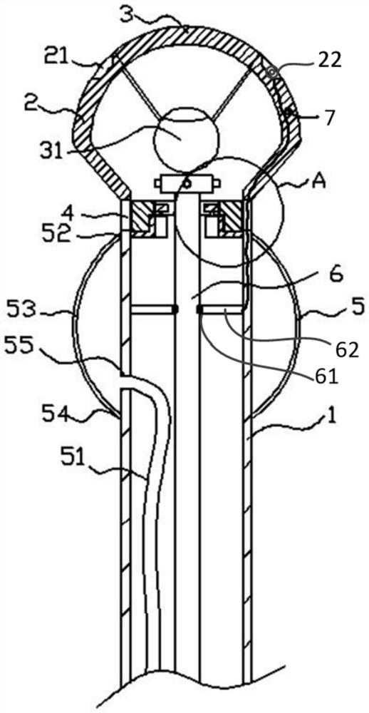
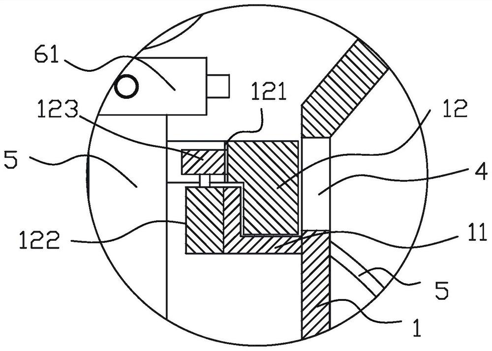
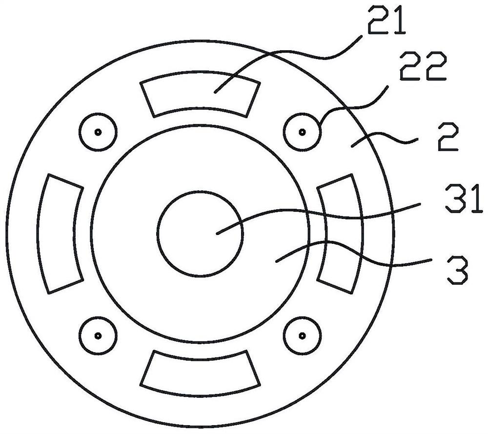
[0027] In order to make the objects, technical solutions, and advantages of the present invention more clearly, the technical solutions in the embodiments of the present invention will be described in contemplation in the embodiment of the present invention. It is an embodiment of the invention, not all of the embodiments. Components of the embodiments of the present invention described and illustrated in the drawings herein can be arranged and design in various configurations.

[0028] Thus, the following detailed description of the embodiments of the invention in the drawings is not intended to limit the scope of the invention claims, but only the selected embodiments of the present invention are shown. Based on the embodiments in the present invention, all other embodiments obtained without creative labor are not made in the premise of creative labor.

[0029] It should be noted that similar reference numerals and letters represent the similar items in the following figures, an...


PUM


Abstract

The invention relates to the field of medical instruments, in particular to a gynecological examination device which comprises an insertion rod (1). The insertion part (2) is arranged at the top end of the insertion rod (1), the insertion part (2) is provided with a transparent view window (3) and a first camera (31) located in the view window (3), and the size of the insertion part (2) is larger than that of the insertion rod (1); the expansion part (5) is coated outside the insertion rod (1); wherein a transparent circular ring window (4) is formed in the position, between the expansion part (5) and the insertion part (2), of the insertion rod (1), a second camera (41) is arranged in the circular ring window (4), and after the expansion part (5) expands, a concave visual field area for the second camera (41) to shoot is formed between the expansion part (5) and the insertion part (2). According to the gynecological examination device, the inner wall of the vagina can be expanded, wrinkles on the inner wall of the vagina are eliminated, images of the inner wall of the vagina are obtained, and vagina examination can be carried out more conveniently and comprehensively.

Description

Technical field [0001] The present invention relates to the field of medical devices, and in particular, there is a gynecological inspection device. Background technique [0002] During some gynecological diseases inspection or treatment, it is necessary to determine the condition or examination of the treatment by viewing the inner wall of the vagina. During the current practice of clinical practice, it is usually observed with naked eye, or uses the camera directly into the female vaginal observation, but due to the small space in the vaginal, the inner wall is wrinkled, there is a check blind spot in some wrinkles, or more The overall situation of the inner wall of the vaginal is observed very well. [0003] In order to obtain a better inspection effect, usually use the expansion of the acoustic wall to open the interior wall, and then insert the camera deep into the vaginal examination. Although this method solves the problem of inspection of the blind zone in the vaginal wal...

Claims


Application Information

Patent Timeline
IPC IPC(8): A61B1/303A61B1/015A61B1/04A61B1/06A61B1/32
CPCA61B1/303A61B1/04A61B1/32A61B1/06A61B1/015
Inventor 李晓琦吴小华
Owner FUDAN UNIV SHANGHAI CANCER CENT
Why Eureka
Try Eureka