
Medical image labeling method, medium and electronic equipment

A technology of medical imaging and seed pixels, which is applied in the field of image analysis and can solve problems such as the inability to realize 3D annotation.

Pending Publication Date: 2021-12-31
杭州普健医疗科技有限公司
View PDF13 Cites 0 Cited by
  • Summary
  • Abstract
  • Description
  • Claims
  • Application Information

AI Technical Summary

Problems solved by technology

[0003] In view of the shortcomings of the prior art described above, the purpose of the present invention is to provide a medical image labeling method, medium and electronic equipment to solve the problem that three-dimensional labeling cannot be achieved in the prior art

Method used


Image

  • Medical image labeling method, medium and electronic equipment
  • Medical image labeling method, medium and electronic equipment
  • Medical image labeling method, medium and electronic equipment

Examples


Embodiment Construction

[0061]Embodiments of the present invention are described below through specific examples, and those skilled in the art can easily understand other advantages and effects of the present invention from the content disclosed in this specification. The present invention can also be implemented or applied through other different specific implementation modes, and various modifications or changes can be made to the details in this specification based on different viewpoints and applications without departing from the spirit of the present invention. It should be noted that, in the case of no conflict, the following embodiments and features in the embodiments can be combined with each other.

[0062] It should be noted that the diagrams provided in the following embodiments are only schematically illustrating the basic ideas of the present invention, and only the components related to the present invention are shown in the diagrams rather than the number, shape and size of the compone...


PUM


Abstract

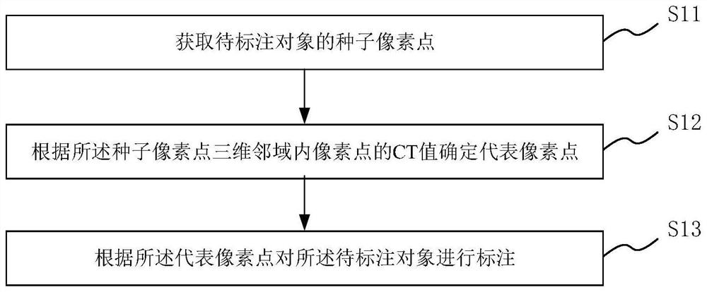
The invention provides a medical image labeling method, a medium and electronic equipment. The medical image labeling method comprises the steps of obtaining seed pixel points of a to-be-labeled object; determining representative pixel points according to the CT values of the pixel points in the three-dimensional neighborhood of the seed pixel points; and labeling the object to be labeled according to the representative pixel points. The medical image labeling method can realize three-dimensional labeling of the to-be-labeled object.

Description

technical field [0001] The invention belongs to the field of image analysis and relates to an image labeling method, in particular to a medical image labeling method, medium and electronic equipment. Background technique [0002] Medical image-aided diagnosis based on artificial intelligence methods needs to collect a large amount of labeled data to train algorithm models. To this end, it is usually necessary to use interactive image segmentation algorithms to mark the location of tumors or organs from existing cases, and then collect enough data for algorithm model training. However, most tumors or organs are three-dimensional structures, and the existing interactive image segmentation algorithms do not have the function of three-dimensional segmentation, which makes it difficult to achieve three-dimensional annotation of tumors or organs. Contents of the invention [0003] In view of the above-mentioned shortcomings of the prior art, the purpose of the present invention...

Claims


Application Information

Patent Timeline
Patent Type & Authority Applications(China)
IPC IPC(8): G06T7/00G06T19/20G16H30/40
CPCG06T7/0012G06T19/20G16H30/40G06T2207/10081G06T2207/30101G06T2207/30204
Inventor 顾静军邓雅卿丁元陈以宁周公敢
Owner 杭州普健医疗科技有限公司
Learn More