Tumor area capillary segmentation method and device

A tumor area and microvessel technology, applied in the field of image processing, can solve the problems of small surface features, inaccurate segmentation, difficult observation of tumor areas, etc., to achieve the effect of improving accuracy, enhancing feature reuse and feature propagation

Pending Publication Date: 2021-12-28
杭州迪英加科技有限公司
View PDF0 Cites 0 Cited by
  • Summary
  • Abstract
  • Description
  • Claims
  • Application Information

AI Technical Summary

Problems solved by technology

However, these methods are often not accurate enough for the segmentation of microvessels in tumor regions because the microvessel segmentation in tumor regions has smaller surface features compared with tissue regions, and tumor regions are not easy to observe.
In

Method used


Image

  • Tumor area capillary segmentation method and device
  • Tumor area capillary segmentation method and device
  • Tumor area capillary segmentation method and device

Examples


Embodiment Construction

[0037] In order to make the purpose, technical solution and advantages of the present application clearer, the present application will be further described in detail below in conjunction with the accompanying drawings and embodiments. It should be understood that the specific embodiments described here are only used to explain the present application, and are not intended to limit the present application.

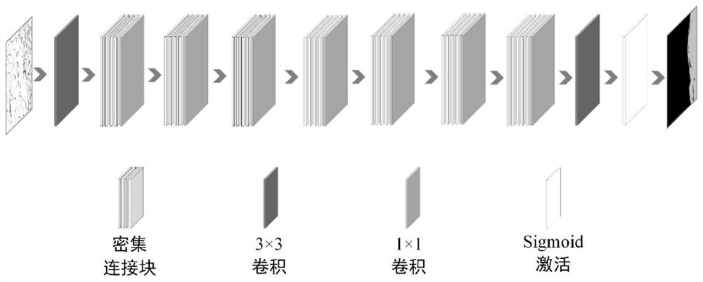
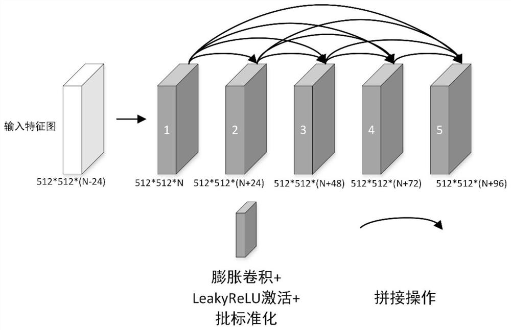
[0038] Fully convolutional neural network (FCN), as one of the typical semantic segmentation networks, has been improved and optimized by many researchers in recent years, and has made good progress in the field of image segmentation. However, these methods adopt a bottom-up decoding method, and the segmentation results obtained by linear interpolation are often rough and inaccurate. On the other hand, in order to obtain more context information, many researchers have added the empty space convolution pooling pyramid (ASPP) to the network, and captured the context informat...


PUM


Abstract

The invention relates to a tumor area capillary segmentation method and device. The method comprises the following steps: acquiring an image block containing a tumor area and microvessels; and inputting the image block into a tumor area microvessel segmentation model to obtain a prediction probability graph of the tumor area and the microvessels, wherein the prediction probability graph comprises a capillary region, a tumor region and a background region, and the tumor area capillary segmentation model comprises two convolution layers with preset scales, a plurality of dense connection blocks and a sigmoid activation module. By adopting the method, the accuracy of tumor area capillary segmentation can be improved.

Description

technical field [0001] The present application relates to the technical field of image processing, in particular to a method and device for segmenting microvessels in tumor regions. Background technique [0002] With the development of image processing technology, automatic recognition and segmentation of target objects through image processing has been applied to various industries. In medical clinical diagnosis, pathologists need to manually segment the microvessels in the tumor area of ​​the breast CD34-stained digital pathological section, and then perform quantitative analysis on the morphological characteristics of each microvessel, and finally classify the patients according to the quantitative analysis results diagnosis. [0003] The existing microvessel segmentation methods in the tumor region of breast CD34 images can often roughly segment the microvessels in the slice. However, these methods are often not accurate enough for the segmentation of microvessels in t...

Claims


Application Information

Patent Timeline
IPC IPC(8): G06T7/194G06T7/11G06T3/40G06N3/04
CPCG06T7/194G06T7/11G06T3/4038G06T2207/30101G06T2207/30096G06T2207/30068G06T2200/32G06N3/048G06N3/045
Inventor 亢宇鑫杨林崔磊
Owner 杭州迪英加科技有限公司
Why Eureka
Try Eureka