
Endoscope image processing method and device, electronic equipment and storage medium

An image processing and endoscopy technology, applied in the field of image processing, can solve the problems of artificial intelligence system assisted diagnosis and treatment resistance and high equipment procurement cost

Pending Publication Date: 2021-04-27
重庆金山医疗技术研究院有限公司
View PDF6 Cites 3 Cited by
  • Summary
  • Abstract
  • Description
  • Claims
  • Application Information

AI Technical Summary

Problems solved by technology

However, medical artificial intelligence systems often require the cooperation of strong GPU (Graphics Processing Unit, graphics processing unit) hardware or professional high-performance computing chips and other hardware, and the equipment procurement costs of these hardware are relatively high, and grassroots hospitals seldom purchase them. It has caused great resistance to promoting the auxiliary diagnosis and treatment of artificial intelligence systems in grassroots hospitals

Method used


Image

  • Endoscope image processing method and device, electronic equipment and storage medium
  • Endoscope image processing method and device, electronic equipment and storage medium
  • Endoscope image processing method and device, electronic equipment and storage medium

Examples


Embodiment Construction

[0043] In order to make the purposes, technical solutions and advantages of the embodiments of the present application clearer, the technical solutions in the embodiments of the present application will be clearly and completely described below in conjunction with the drawings in the embodiments of the present application. Obviously, the described embodiments It is only a part of the embodiments of the present application, but not all the embodiments. Based on the embodiments in this application, all other embodiments obtained by persons of ordinary skill in the art without making creative efforts belong to the scope of protection of this application.

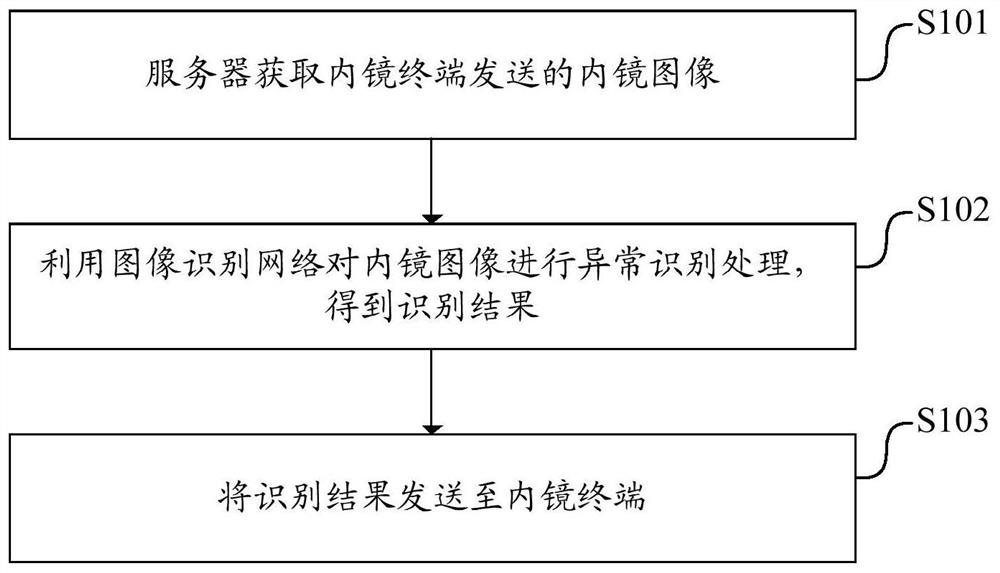
[0044] Please refer to figure 1 , figure 1 It is a flow chart of an endoscopic image processing method provided by the embodiment of this application. The method includes:

[0045] S101: The cloud acquires the endoscopic image sent by the endoscopic terminal.

[0046] All or part of the steps in the endoscopic image process...


PUM


Abstract

The invention discloses an endoscopic image processing method and device, electronic equipment and storage medium. The method comprises the steps of obtaining an endoscopic image sent by an endoscopic terminal by a cloud; performing anomaly identification processing on the endoscopic image by using an image identification network to obtain an identification result; sending the identification result to an endoscope terminal. According to the method, the endoscope terminal does not need to process the endoscope image, so that the hardware requirement on the endoscope terminal can be reduced, the cost of the endoscope terminal is reduced, and the endoscope terminal can be widely applied; through the cooperation of the cloud and the plurality of endoscope terminals, the endoscope terminals can be widely applied, the film reading workload is effectively reduced, the efficiency is improved, and the auxiliary diagnosis and treatment of an artificial intelligence system in primary hospitals can be effectively promoted.

Description

technical field [0001] The present application relates to the technical field of image processing, and in particular to an endoscopic image processing method, an endoscopic image processing device, electronic equipment, and a computer-readable storage medium. Background technique [0002] The incidence of digestive tract diseases is relatively high in the world, especially with the weakening of various body functions, middle-aged and elderly people have become a group with a higher incidence of digestive tract diseases. In my country, most of the inspections for digestive tract diseases are completed in primary hospitals. Capsule endoscopy is convenient and has low physical requirements for middle-aged and elderly people. It is suitable for the examination of any gastrointestinal symptoms. It has no side effects and is non-invasive. However, in the process of digestive tract examination with capsule endoscopy, a large amount of image data will be generated, ranging from a f...

Claims


Application Information

Patent Timeline
Patent Type & Authority Applications(China)
IPC IPC(8): G06T7/00G16H30/40G06F11/14G06F21/60
CPCG06T7/0012G16H30/40G06F11/1448G06F21/602G06T2207/10068G06T2207/30028
Inventor 黄访廖静吴吉芳
Owner 重庆金山医疗技术研究院有限公司
Learn More