Vessel Segmentation Method of Fundus Image Based on Shared Decoder and Residual Tower Structure

A fundus image and tower structure technology, which is applied in the field of image processing, can solve the problems of weak contrast of fundus images and uneven distribution of blood vessel diameters, and achieve the effect of improving the segmentation effect.

Active Publication Date: 2022-03-08
SUN YAT SEN UNIV
View PDF5 Cites 0 Cited by
  • Summary
  • Abstract
  • Description
  • Claims
  • Application Information

AI Technical Summary

Problems solved by technology

[0007] In order to overcome the problems of uneven distribution of blood vessel calibers and weak contrast of fundus images in the prior art when segmenting fundus image blood vessels, the present invention provides a fundus image blood vessel segmentation method based on a shared decoder and a residual tower structure

Method used


Image

  • Vessel Segmentation Method of Fundus Image Based on Shared Decoder and Residual Tower Structure
  • Vessel Segmentation Method of Fundus Image Based on Shared Decoder and Residual Tower Structure
  • Vessel Segmentation Method of Fundus Image Based on Shared Decoder and Residual Tower Structure

Examples


Embodiment 1

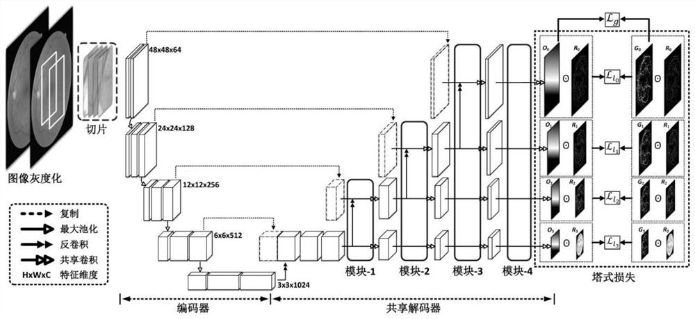
[0077] Such as figure 1 As shown, the fundus image blood vessel segmentation method based on the shared decoder and the residual tower structure, the method is implemented by a processing module, and the processing module includes: a data input module, a residual tower module, an encoding module, and a shared decoding module , loss module, data output module, among them, such as figure 2 As shown, the encoding module and the shared decoding module constitute a U-shaped network with a total of 2L layers (in a specific embodiment, L=5, 2L=10), as image 3 Shown is a residual tower diagram, the method includes the following steps:

[0078] S1: The data input module receives the labeled training data set and the test data set to be divided, and performs slice preprocessing respectively to obtain training data set image blocks and test data set image blocks;

[0079] More specifically, step S1 includes:

[0080] S101: Input the two-dimensional RGB fundus image in the training d...


PUM


Abstract

The invention discloses a fundus image blood vessel segmentation method based on a shared decoder and a residual tower structure. The method comprises the following steps: obtaining an image block of a training data set and an image block of a test data set through a data input module; The tower module obtains the residual tower sequence; the multi-level semantic features are obtained through the encoding module; the multi-level probability map is obtained through the shared decoding module; the multi-scale label, the residual tower sequence, and the probability map obtained by the shared decoder are constructed The total loss of the model is formed, and PyTorch is used for gradient optimization to train the parameters in the encoding module and the shared decoding module; the image blocks of the test data set are sequentially input into the trained encoding module and the shared decoding module to obtain a probability map, and the obtained probability map And stitching, binarization processing to get the final segmentation results. The invention solves the problems of uneven distribution of blood vessel calibers and weak contrast of fundus images.

Description

technical field [0001] The invention relates to the technical field of image processing, and more specifically, to a fundus image blood vessel segmentation method based on a shared decoder and a residual tower structure. Background technique [0002] Accurate segmentation of retinal vessels plays a key role in the diagnosis of diabetic retinopathy, age-related macular degeneration, glaucoma and other ophthalmic diseases. The purpose of this technology is to classify fundus images at the pixel level, that is, to determine whether each pixel is a retinal blood vessel. [0003] For the segmentation of retinal blood vessels, the current mainstream technologies include U-Net and its improved methods. A U-shaped network consists of an encoder and a decoder connected in series. In order to improve the segmentation effect of U-shaped network, the main improvement methods are multi-modular network method (MS-NFN) and double-pass encoded U-shaped network (DEU-Net). [0004] Multi-m...

Claims


Application Information

Patent Timeline
Patent Type & Authority Patents(China)
IPC IPC(8): G06T7/00G06T7/10G06T3/40
Inventor 任传贤许耿鑫
Owner SUN YAT SEN UNIV
Why Eureka
Try Eureka