
Left ventricle segmentation method and device and electronic equipment

A left ventricle and heart technology, applied in the field of medical images, can solve problems such as difficult left ventricle, accurate segmentation, etc.

Active Publication Date: 2021-02-02
SAINUO WEISHENG SCI & TECH BEIJING
View PDF15 Cites 3 Cited by
  • Summary
  • Abstract
  • Description
  • Claims
  • Application Information

AI Technical Summary

Problems solved by technology

[0012] This application provides a left ventricle segmentation method, device and electronic equipment to solve the technical problem in the related art that it is difficult to accurately segment the left ventricle

Method used


Image

  • Left ventricle segmentation method and device and electronic equipment
  • Left ventricle segmentation method and device and electronic equipment
  • Left ventricle segmentation method and device and electronic equipment

Examples


Embodiment Construction

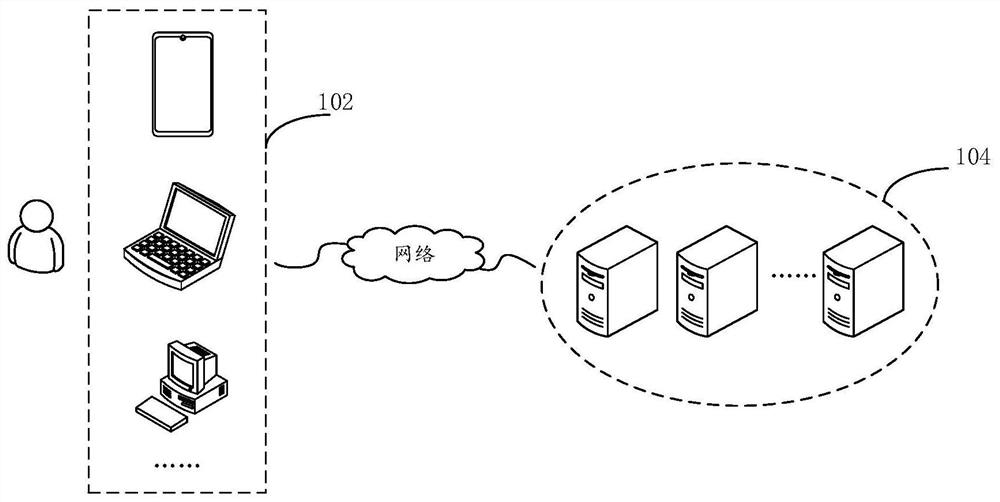
[0034] In order to make those skilled in the art better understand the solutions of the present application, the technical solutions in the embodiments of the present application will be clearly and completely described below with reference to the accompanying drawings in the embodiments of the present application. Obviously, the described embodiments are only The embodiments are part of the present application, but not all of the embodiments. Based on the embodiments in the present application, all other embodiments obtained by those of ordinary skill in the art without creative work shall fall within the scope of protection of the present application.

[0035] It should be noted that the terms "first", "second", etc. in the description and claims of the present application and the above drawings are used to distinguish similar objects, and are not necessarily used to describe a specific sequence or sequence. It is to be understood that data so used may be interchanged under ...


PUM


Abstract

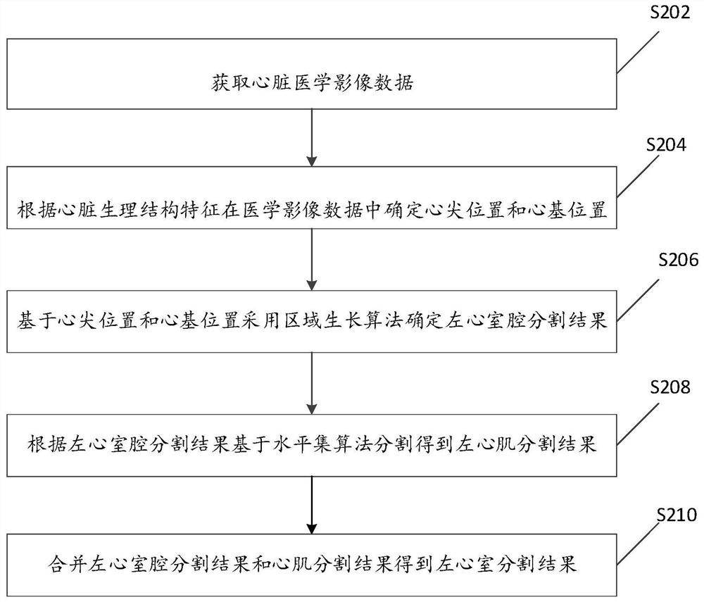
The invention provides a left ventricle segmentation method and device and electronic equipment, and the method comprises the steps of obtaining cardiac medical image data; determining a cardiac apexposition and a cardiac base position in the medical image data according to the heart physiological structure characteristics; determining a left ventricular cavity segmentation result by adopting a region growing algorithm based on the cardiac apex position and the cardiac base position; according to the left ventricular cavity segmentation result, performing segmentation based on a level set algorithm to obtain a left myocardial segmentation result; and combining the left ventricular cavity segmentation result and the left myocardial segmentation result to obtain a left ventricular segmentation result. As the apex and the heart base are located in the ventricle, an accurate left ventricular cavity segmentation result can be obtained by adopting a region growing method based on the apex and the heart base, and due to the fact that the left ventricular cavity and the left myocardium are large in gray scale difference and obvious in gray scale gradient change, accurate left myocardium segmentation is conducted through a level set algorithm on the basis of the left ventricular cavity segmentation result, and the evolution speed is fast.

Description

technical field [0001] The present application relates to the technical field of medical images, and in particular, to a left ventricle segmentation method, device and electronic device. Background technique [0002] Heart disease has become the number one killer of human health in recent years. How to effectively and accurately diagnose cardiovascular disease has become the number one issue for medical workers. From the perspective of cardiac mechanism, the left ventricle, which is responsible for supplying blood to various organs in the body, is the most important part of the heart. Therefore, the abnormality of its shape and movement is regarded as an important basis for the evaluation of cardiac disease. With the development of medical imaging technology, non-invasive analysis of cardiac function based on the left ventricle has become an important auxiliary method for clinical diagnosis of cardiac diseases. [0003] At present, there are many imaging methods for cardi...

Claims


Application Information

Patent Timeline
IPC IPC(8): G06T7/00G06T7/11
CPCG06T7/0012G06T2207/30048G06T7/11
Inventor 黄艳孙奇马双袁玉亮王金娇
Owner SAINUO WEISHENG SCI & TECH BEIJING
Learn More