Pretreatment solution, kit and pretreatment method for fluorescence in situ hybridization of cell samples
A technique of fluorescence in situ hybridization and pretreatment liquid, which is applied in the fields of biochemical equipment and methods, and the determination/inspection of microorganisms, which can solve the problems of decreased activity and incomplete enzymatic digestion.
- Summary
- Abstract
- Description
- Claims
- Application Information
AI Technical Summary
Problems solved by technology
Method used
Image

Examples
Embodiment 1
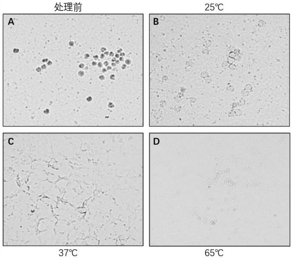
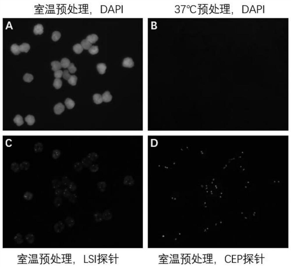
[0050] After cytological preparation with normal human blood, pretreatment was carried out according to the method of the present invention (the steps shown in Table 1). postnatal results. After pretreatment, observe under a bright-field microscope whether there are still cell outlines. Then FISH hybridization and DAPI staining were performed. Observe under a fluorescent microscope whether there are DAPI-stained nuclei, and the signal points of the orange gene-specific probe and the green chromosome CEP probe.
[0051] The test results are shown in Table 1 and figure 1 with figure 2 As shown, among them, figure 1 It shows the profile of normal human blood cell droplet under bright field microscope after pretreatment with 100mM NaOH, 0.5% Triton x-100 solution at different temperatures; figure 2 Shown is the FISH signal of the normal human blood cell droplet under the fluorescence microscope after pretreatment with 100mM NaOH, 0.5% Triton x-100 solution.
[0052] figur...
Embodiment 2
[0057] In Example 2, the same blood cell sample as in Example 1 was used for fluorescence in situ hybridization, except that the steps shown in Table 2 were used for pretreatment, and more experimental conditions were tried. The test results are shown in Table 2.
[0058] Table 2:
[0059]
[0060]
[0061]
[0062]It can be seen from Table 2 that after the blood cells are treated with the pretreatment solution containing 15-150 mM lye and 0.1%-1% non-ionic surfactant, FISH signals can be detected. Wherein, the fluorescence in situ detection effect of the blood cells after the pretreatment solution containing 100mM KOH and 0.5% NP-40 is equivalent to the treatment effect of the pretreatment solution containing 100mM NaOH and 0.5% TritonX-100 in Example 1, in The fluorescence images at 25°C are relatively clear. The second is the pretreatment solution containing 15mM NaOH and 0.8% Triton X-100, and the pretreatment solution containing 150mM NaOH and 0.3% Triton X-100...
Embodiment 3
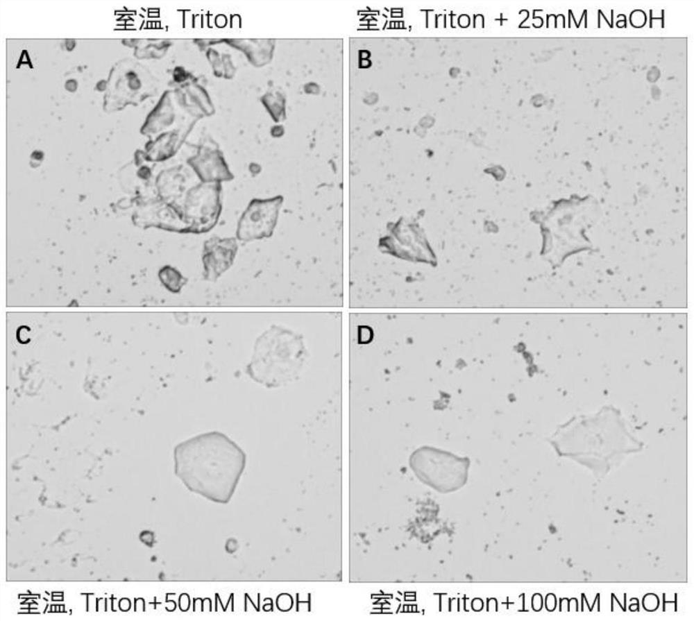
[0064] According to the conventional method, exfoliated cells in the urine of normal people were used for preparation. After sheeting, pretreatment was carried out according to the method of the present invention (shown in Table 3). In this example, different treatment fluid formulations were tested (as shown in Table 3 below). Observation by bright field microscopy was performed after pretreatment. Then, FISH hybridization and DAPI staining were performed according to conventional methods, and observed under a fluorescent microscope.
[0065] table 3:
[0066]
[0067] The test results are shown in Table 3 and image 3 and Figure 4 As shown, among them, image 3 It shows the profile of the exfoliated cell droplet in normal human urine under bright field microscope after the solution of different formulations pretreated the exfoliated cells in urine at room temperature; Figure 4 It shows the FISH signal under the fluorescence microscope after the normal human urine ...
PUM
 Abstract
Description
Claims
Application Information
 - R&D
- Intellectual Property
- Life Sciences
- Materials
- Tech Scout
- Unparalleled Data Quality
- Higher Quality Content
- 60% Fewer Hallucinations
Browse by: Latest US Patents, China's latest patents, Technical Efficacy Thesaurus, Application Domain, Technology Topic, Popular Technical Reports.
© 2025 PatSnap. All rights reserved.Legal|Privacy policy|Modern Slavery Act Transparency Statement|Sitemap|About US| Contact US: help@patsnap.com