Image display method and device, angiography device and storage medium
An angiography and image display technology, applied in cardiac catheterization, medical science, and equipment for radiological diagnosis, etc., to achieve the effects of reducing frequency and radiation dose, increasing service life, and improving diagnostic efficiency
- Summary
- Abstract
- Description
- Claims
- Application Information
AI Technical Summary
Problems solved by technology
Method used
Image
Examples
Embodiment 1
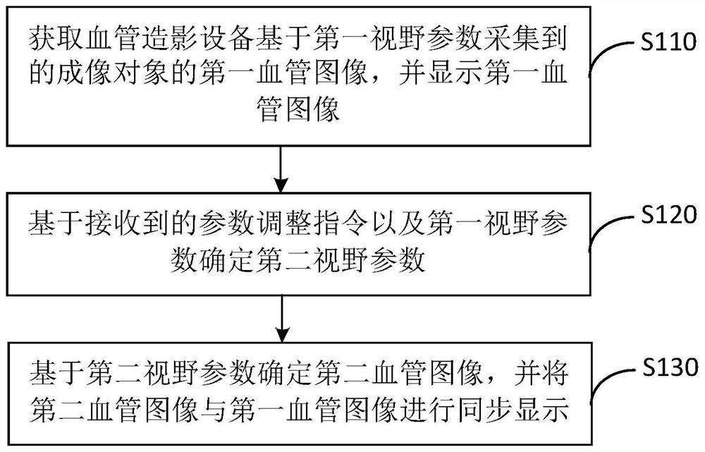
[0026] figure 1 It is a flow chart of an image display method provided by Embodiment 1 of the present invention. This embodiment is applicable to the situation where angiography equipment is used for imaging and the imaging result is displayed. This method can be executed by an image display device. The device can be realized by means of software and / or hardware, and the device can be configured in angiography equipment. Specifically include the following steps:
[0027] S110. Acquire a first blood vessel image of an imaging object acquired by an angiography device based on a first field of view parameter, and display the first blood vessel image.
[0028] Angiography is a technique for visualizing blood vessels in X-ray sequence images. By injecting contrast agents into blood vessels, since X-rays cannot penetrate contrast agents, angiography uses X-rays to scan and image the measured site. Angiography The agent makes the blood vessels in the measured part and other tissues...
Embodiment 2
[0044] figure 2 It is a flow chart of an image display method provided in Embodiment 2 of the present invention. This embodiment is further refined on the basis of the above embodiments. Optionally, the second blood vessel image and the first Synchronously displaying a blood vessel image includes: when the first field of view parameter corresponding to the first blood vessel image changes, acquiring an updated first blood vessel image acquired by the angiography device based on the changed first field of view parameter; The first visual field parameter and the parameter adjustment instruction determine to update the second visual field parameter, and display the updated second blood vessel image determined based on the updated second visual field parameter and the updated first blood vessel image synchronously.
[0045] The specific implementation steps of this embodiment include:
[0046] S210. Acquire a first blood vessel image of an imaging object acquired by an angiograp...
Embodiment 3
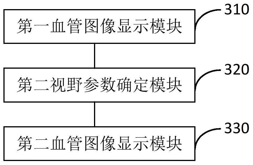
[0065] image 3 It is a schematic diagram of an image display device provided in Embodiment 3 of the present invention. This embodiment is applicable to the case of using angiographic equipment to perform imaging and display the imaging results. The apparatus can be implemented in software and / or hardware, and the apparatus can be configured in the angiographic equipment. The image display device includes: a first blood vessel image display module 310 , a second visual field parameter determination module 320 and a second blood vessel image display module 330 .
[0066] Wherein, the first blood vessel image display module 310 is configured to acquire the first blood vessel image of the imaging object collected by the angiography device based on the first field of view parameter, and display the first blood vessel image;
[0067] The second field of view parameter determination module 320 is configured to determine a second field of view parameter based on the received paramet...
PUM
 Abstract
Description
Claims
Application Information
 - R&D
- Intellectual Property
- Life Sciences
- Materials
- Tech Scout
- Unparalleled Data Quality
- Higher Quality Content
- 60% Fewer Hallucinations
Browse by: Latest US Patents, China's latest patents, Technical Efficacy Thesaurus, Application Domain, Technology Topic, Popular Technical Reports.
© 2025 PatSnap. All rights reserved.Legal|Privacy policy|Modern Slavery Act Transparency Statement|Sitemap|About US| Contact US: help@patsnap.com


