
Three-dimensional continuous culture method of porcine mammary epithelial cells

A technology of epithelial cells and culture methods, which is applied in the field of three-dimensional continuous culture of porcine mammary gland epithelial cells, and achieves the effect of time-saving and simple operation

Pending Publication Date: 2020-05-01
ANIMAL SCI RES INST GUANGDONG ACADEMY OF AGRI SCI
View PDF1 Cites 6 Cited by
  • Summary
  • Abstract
  • Description
  • Claims
  • Application Information

AI Technical Summary

Problems solved by technology

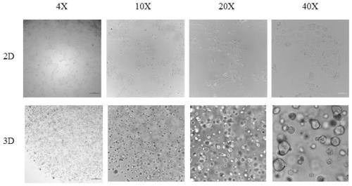
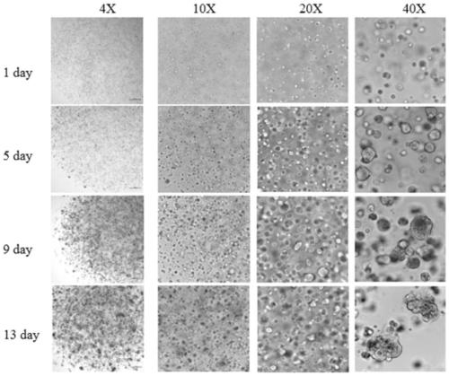
[0004] At present, there are few reports on the combination of porcine mammary epithelial cells and three-dimensional cell culture methods. In order to improve the defects of the traditional monolayer cell culture method in mammary epithelial cell culture, a three-dimensional continuous culture method of porcine mammary epithelial cells is provided. For porcine mammary gland The study of epithelial cells is of great significance

Method used


Image

  • Three-dimensional continuous culture method of porcine mammary epithelial cells
  • Three-dimensional continuous culture method of porcine mammary epithelial cells

Examples


Embodiment 1

[0029] A three-dimensional continuous culture method for porcine mammary gland epithelial cells, comprising the following steps:

[0030] (1) Separation and extraction of primary porcine mammary epithelial cells before the 10th generation;

[0031] (2) Digest porcine mammary gland epithelial cells into single cells with trypsin, centrifuge at 1000rpm for 5min, discard the supernatant, add 1mL ordinary medium to resuspend, adjust the cell concentration to 1×10 6 cells / mL, after centrifugation at 800rpm for 5min, discard the supernatant and add 1mL experimental Matrigel to resuspend, mix well and store on ice for later use;

[0032] (3) Use a 200 μL pipette tip to absorb the above suspension, drop 30 μL / drop into a six-well plate, 8 drops per well, turn it upside down after dropping, and incubate in a 37°C carbon dioxide incubator for 30 minutes to solidify;

[0033] (4) Add 2 mL of three-dimensional medium to each well of a six-well plate, and continue culturing for 14 days, a...

Embodiment 2

[0039] A three-dimensional continuous culture method for porcine mammary gland epithelial cells, comprising the following steps:

[0040] (1) Separation and extraction of cells from the primary epithelium of porcine mammary glands before the 10th passage;

[0041] (2) Digest porcine mammary gland epithelial cells into single cells with trypsin, centrifuge at 1000rpm for 4min, discard the supernatant, add 1mL ordinary medium to resuspend, adjust the cell concentration to 5×10 6 cells / mL, after centrifugation at 800rpm for 6min, discard the supernatant and add 1mL experimental Matrigel to resuspend, mix well and store on ice for later use;

[0042] (3) Use a 200 μL pipette tip to absorb the suspension obtained in step (2), drop 20 μL / drop into a six-well plate, 8 drops per well, and incubate for 40 minutes in a 37°C carbon dioxide incubator after dropping. its solidification;

[0043] (4) Add 2 mL of three-dimensional medium to each well of a six-well plate, and continue cultu...

Embodiment 3

[0049] A three-dimensional continuous culture method for porcine mammary gland epithelial cells, comprising the following steps:

[0050] (1) Separation and extraction of primary porcine mammary epithelial cells before the 10th generation;

[0051] (2) Digest porcine mammary gland epithelial cells into single cells with trypsin, centrifuge at 1000rpm for 6min, discard the supernatant, add 1mL ordinary medium to resuspend, adjust the cell concentration to 5×10 6 cells / mL, after centrifugation at 800rpm for 4min, discard the supernatant and add 1mL experimental Matrigel to resuspend, mix well and store on ice for later use;

[0052] (3) Use a 200 μL pipette tip to absorb the suspension obtained in step (2), drop 40 μL / drop into a six-well plate, 7 drops per well, and incubate for 35 minutes in a 37°C carbon dioxide incubator after dropping. its solidification;

[0053] (4) Add 2 mL of three-dimensional medium to each well of a six-well plate, and continue culturing for 16 days...


PUM


Abstract

The present invention relates to a three-dimensional continuous culture method of porcine mammary epithelial cells and belongs to the field of cell culture methods. The three-dimensional continuous culture method comprises the following specific steps: (1) separating and extracting primary porcine mammary epithelial cells and selecting cells before 10th generation; (2) after using trypsin to conduct digestion into singe cells, conducting centrifugation, discarding supernatant, conducting re-suspending in a common culture medium, adjusting cell concentration to 1x10<6>-5x10<6> cells / mL, taking1 mL of the cells, then conducting centrifugation again, discarding supernatant, conducting re-suspending with Matrigel for experimental use, and conducting even mixing for standby application on ice;(3) drawing an appropriate amount of the above suspension, dropping 7-8 drops into each well of a six-well plate, conducting inversion after the dropping, and conducting placing in a 37 DEG C incubator for incubation for 30-40 min for coagulation; (4) adding 2 mL of a three-dimensional culture medium per well, conducting continuous culture for 12-16 days, and changing the three-dimensional culture medium once every 2-3 days according to color of the culture medium; and (5) removing the culture medium, conducting washing with D-PBS for three times, adding 2 mL of trypsin to each well, using apipette tip for blowing off, conducting culture at 37 DEG C, conducting blowing off to conducting even mixing every other 2-4 min until single cells are obtained, conducting neutralization digestion and centrifugation at 4 DEG C, removing supernatant, and conducting passage according to the step (2) to step (4).

Description

technical field [0001] The invention relates to the field of cell culture methods, in particular to a three-dimensional continuous culture method for porcine mammary gland epithelial cells. Background technique [0002] Mammary gland epithelial cells are the main cells that make up the mammary gland and have a special function of milk secretion. Many components of milk can only be synthesized by mammary gland epithelial cells. The study of mammary gland cells began in the 1970s. It has become a hot spot to cultivate primary mammary gland epithelial cells at the cellular level through effective cell culture methods and conduct passages to study mammary gland growth, development, lactating cells or molecular biological mechanisms. . At present, there are many studies on human, mouse, and bovine mammary epithelial cells, but there are few reports on porcine mammary epithelial cells. [0003] As the most core and basic technology in medical biotechnology, cell technology has b...

Claims


Application Information

Patent Timeline
IPC IPC(8): C12N5/071
CPCC12N5/0631C12N2500/25C12N2500/32C12N2501/11C12N2501/155C12N2501/39C12N2501/405C12N2501/415C12N2513/00C12N2533/90
Inventor 肖昊查翠芳王丽杨雪芬蒋宗勇
Owner ANIMAL SCI RES INST GUANGDONG ACADEMY OF AGRI SCI
Learn More