
A radiation-free percutaneous spine localization method based on optical scanning automatic contour segmentation and matching

An optical scanning and contouring technology, applied in the fields of medical technology and computer image processing, can solve the problems of inability to achieve uniform size, inconsistent coordinate starting points, no anatomical landmarks, etc. Intuitive results

Active Publication Date: 2021-04-13
ZHEJIANG UNIV
View PDF15 Cites 0 Cited by
  • Summary
  • Abstract
  • Description
  • Claims
  • Application Information

AI Technical Summary

Problems solved by technology

However, in the medical field, because the image is more complex and contains more signals, and the information inside the human body contour is the focus of clinical treatment, the optical scanning technology that can only recognize the human body contour alone cannot achieve perspective, so Difficult to use in clinical treatment
In recent years, with the maturity of image fusion technology, there have been many technologies that try to fuse optical scanning images and medical scanning images, but these technologies are often difficult to achieve automatic matching, and they can only find fixed and prominent feature points manually. The parts of the human body (such as the nose and ears of the human body) are used as the target points for contour matching. The reason lies in the complexity and density inhomogeneity of human medical image signals. The 3D point cloud generated based on medical images cannot achieve uniform and equal size. , it is difficult to match the homogeneous 3D point cloud generated based on optical scanning
In addition, the inconsistency of the three-dimensional coordinate ratio of the human medical image and the optical scanning image, and the inconsistency of the starting point of the coordinates will also cause the matching image to be rotated, embedded, shifted, etc.
[0007] The skin on the back of the human body is highly mobile, has no fixed surface shape, and has no characteristic anatomical landmarks. Its appearance can change with the body's height, fat, and thinness, and can also change with the change of body position, so it is difficult to optically scan images and clinical medical images for matching

Method used


Image

  • A radiation-free percutaneous spine localization method based on optical scanning automatic contour segmentation and matching
  • A radiation-free percutaneous spine localization method based on optical scanning automatic contour segmentation and matching
  • A radiation-free percutaneous spine localization method based on optical scanning automatic contour segmentation and matching

Examples


Embodiment Construction

[0050] The present invention will be further described below in conjunction with the accompanying drawings.

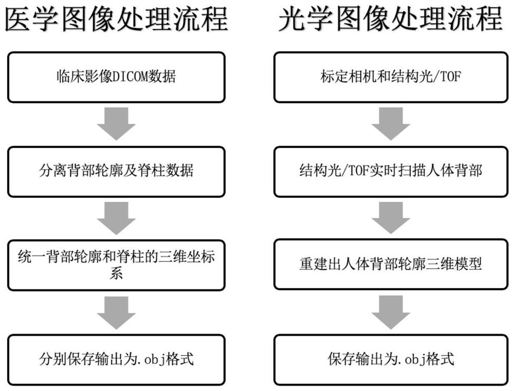
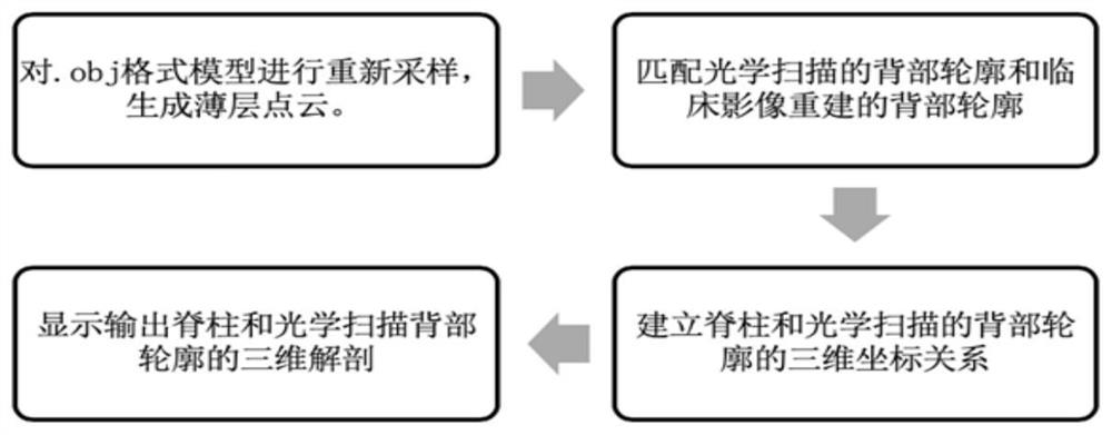
[0051] Such as figure 1 and 2 As shown, the process involved in the method of the present invention includes: a medical image processing process and an optical image processing process.

[0052] 1. Medical image processing flow:

[0053] 1.1: Obtain clinical image DICOMs data (taking CT as an example) and input it into the image processing system (3).

[0054] 1.2: Separate and extract the CT value of the human spine through a fixed CT threshold (Threshold) range. The threshold must meet the requirement of only displaying bone density (>400Hu) without any other tissue density. After extraction, the bone density is used to reconstruct the output A 3D model of the human spine and save it in .obj format.

[0055] 1.3: Separate and extract the CT value of the human body contour through a fixed CT threshold (Threshold) range, which must meet the requirements of not cont...


PUM


Abstract

The invention discloses a radiation-free percutaneous spine positioning method based on optical scanning automatic contour segmentation and matching. The clinical image data (CT) is imported into the clinical image data processing system to segment and reconstruct the three-dimensional model of the back contour and spine of the human body. Through the structure Light / TOF (Time of flight) optical scanning equipment scans and reconstructs the 3D model of the human back contour, and finally, after the matching calculation of the 3D image processing system, it displays the 3D coordinate relationship between the output spine model and the optically scanned human back contour model. The method of the invention is based on an optical scanning device, realizes matching and positioning of a spine model and an optically scanned human back contour model, and has the advantages of no radiation, less space occupation, fast scanning speed, intuitive display results, and high repeatability.

Description

technical field [0001] The invention relates to the fields of medical technology and computer image processing, and relates to a radiation-free percutaneous spine positioning method based on optical scanning automatic contour segmentation and matching. Background technique [0002] At present, percutaneous minimally invasive spinal surgery has been applied to intervertebral discectomy, laminectomy decompression, foraminotomy, cyst and tumor resection, etc. Its advantages lie in small trauma and can protect the spine and its surrounding areas to the greatest extent. anatomy. [0003] Percutaneous endoscopic surgery is a representative of percutaneous minimally invasive spinal surgery. Its trauma is small and intuitive. The display system is common with other surgical endoscopic platforms. The economic investment is not high, and it has good clinical application prospects. There are higher requirements for intraoperative positioning and puncture. Usually, the spine surgeon n...

Claims


Application Information

Patent Timeline
Patent Type & Authority Patents(China)
IPC IPC(8): A61B34/10A61B34/20
CPCA61B34/10A61B34/20A61B2034/105A61B2034/107A61B2034/108A61B2034/2055
Inventor 江晓航朱永坚
Owner ZHEJIANG UNIV
Learn More