Key CT technology of lung carcinoma in-situ identification method

An identification method, CT technology, applied in the fields of radiological diagnostic equipment, medical science, diagnosis, etc., can solve the problems that have not been reported, and achieve the effect of clear images

Inactive Publication Date: 2020-01-10
SHANGHAI PULMONARY HOSPITAL
View PDF6 Cites 1 Cited by
  • Summary
  • Abstract
  • Description
  • Claims
  • Application Information

AI Technical Summary

Problems solved by technology

[0010] But about the key CT technology of a kind of lung carcinoma in situ identification method of the present invention, there is no report yet

Method used


Image

  • Key CT technology of lung carcinoma in-situ identification method

Examples


Embodiment 1

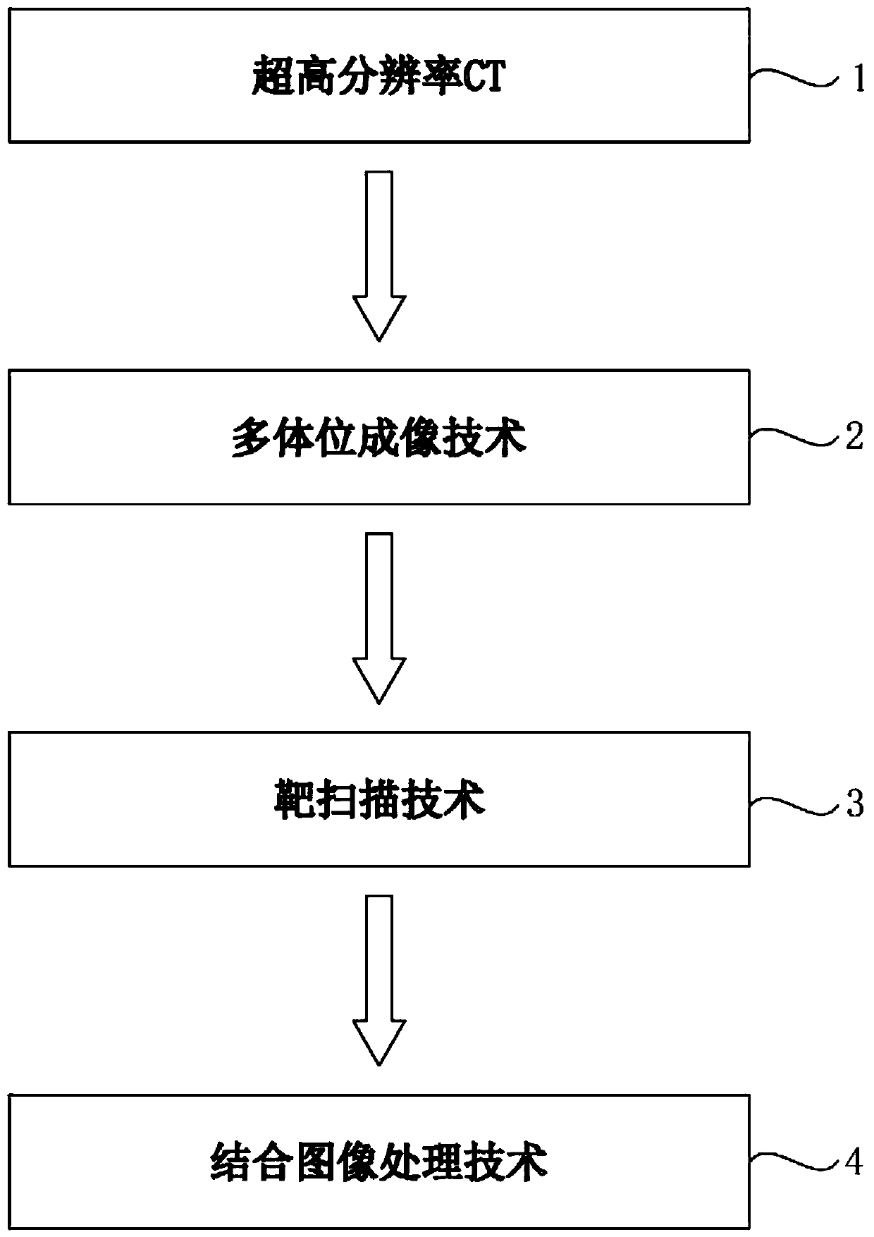
[0030] Please refer to the attached figure 1 , Attached figure 1 It is a schematic diagram of the plane module of the key CT technology of a method for identifying lung carcinoma in situ in this embodiment. The key CT technologies of the lung carcinoma in situ recognition method include ultra-high resolution CT1, multi-position imaging technology 2, target scanning technology 3, combined image processing technology 4;

[0031] The said ultra-high resolution CT1 is an ultra-high resolution CT based on a 1024×1024 matrix, and its resolution is 4 times higher than that of a conventional CT based on a 512×512 matrix;

[0032] The multi-position imaging technique 2 is supine, prone, left-side, and right-side imaging;

[0033] The described target scanning technology 3 uses a small field of view (FOV) under the same matrix to reduce pixels and improve spatial resolution;

[0034] The combined image processing technology 4 is to analyze the characteristic imaging performance of ground glas...

Embodiment 2

[0036] The specific identification method and process of the key CT technology of the lung carcinoma in situ identification method:

[0037] Ultra-high resolution CT based on a 1024×1024 matrix is ​​used; when the matrix is ​​the same, a small field of view (FOV) is used to reduce pixels and improve spatial resolution; and multi-position and multi-angle imaging are used to keep the lesion away On the side of the examination table, the influence of the vascular drop effect is minimized, thereby increasing the image contrast and displaying the lesion more clearly.

[0038] It should be noted that: unlike the original data image obtained by conventional scanning for target reconstruction, target scanning is not a simple geometric enlargement, and the image is clearer.

[0039] Instead, the target scan is based on ultra-high-resolution CT, multi-position imaging, so that the resulting image is clearer, and can clearly show the internal fine structure, density, boundary and surrounding si...


PUM


Abstract

The invention relates to a key CT technology of a lung carcinoma in-situ identification method. The key CT technology of the lung carcinoma in-situ identification method comprises an ultra-high resolution CT, a multi-position imaging technology, a target scanning technology and a combined image processing technology. The key CT technology has the advantages that target reconstruction is carried out unlike on an original data image obtained by conventional scanning, target scanning is not a simple geometric enlargement, and the image is clearer; instead, the target scanning is based on ultra-high resolution CT, multi-position imaging is achieved, the obtained image is enabled to be clearer, and the internal microstructure, density, border and surrounding signs of small nodules can be clearly showed; and moreover, ultra-high resolution combined with the multi-position imaging technology and the target scanning technology can analyze the characteristic imaging findings of ground glass nodules in shape, edge, density, diameter, internal solid components, vascular abnormalities and pleural depression, and early accurate diagnosis is expected to be performed on AAH and AIS and further guide clinical practice.

Description

Technical field [0001] The present invention relates to the technical field of lung cancer in situ identification, and specifically, is a key CT technology of a method for identifying lung cancer in situ. Background technique [0002] Lung cancer is the malignant tumor with the highest incidence and mortality in the world. According to statistics, the pathological type with the highest incidence of lung cancer is lung adenocarcinoma, which accounts for 65.4% of the detected lung cancers. Lung adenocarcinoma is divided into pre-invasive lesions (atypical adenomatous hyperplasia, AAH and adenocarcinoma in situ, adenocarcinoma in situ, AIS), minimally invasive adenocarcinoma (MIA), and invasive adenocarcinoma And four major types of invasive adenocarcinoma variants. Among the four types, the imaging manifestations of pre-invasive lesions, micro-invasive adenocarcinoma, and invasive adenocarcinoma have certain characteristics and are easy to diagnose. However, AAH and AIS are two co...

Claims


Application Information

Patent Timeline
IPC IPC(8): A61B6/03
CPCA61B6/032A61B6/5229
Inventor 孙希文孙珂王斌杨洋
Owner SHANGHAI PULMONARY HOSPITAL
Why Eureka
Try Eureka