A kind of foramen ovale positioning method, device and storage medium
A positioning method and foramen ovale technology, applied in the field of image processing, can solve the problems of inability to guarantee positioning accuracy and reduce positioning efficiency, and achieve the effect of improving positioning accuracy and positioning efficiency
- Summary
- Abstract
- Description
- Claims
- Application Information
AI Technical Summary
Problems solved by technology
Method used
Image
Examples
Embodiment 1
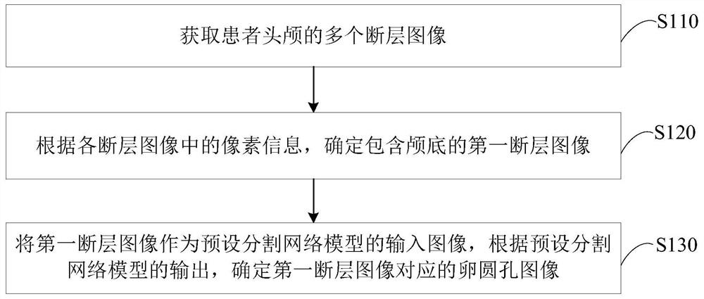
[0027] figure 1 A flow chart of a foramen ovale positioning method provided in the first embodiment of the present invention, this embodiment can be applied to the situation of positioning the foramen ovale in the skull base, and the method can be performed by a foramen ovale positioning device, The apparatus can be implemented in software and / or hardware, and be integrated into a device with image processing functions. The method specifically includes the following steps:
[0028] S110. Acquire multiple tomographic images of the patient's skull.
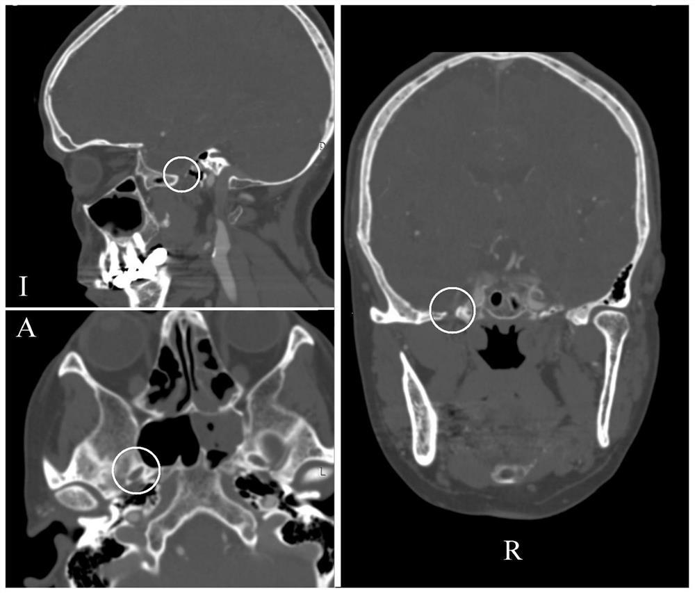
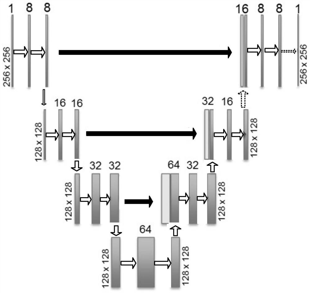
[0029] Wherein, the tomographic image may refer to an image obtained by scanning a patient's head with a scanning device and reconstructing according to the obtained scanning data. The scanning equipment may be, but is not limited to, X-ray photography (X-ray photography), CT (Computed Tomography, electronic computer tomography scanning equipment) and MR (Magnetic Resonance Imaging, magnetic resonance scanning equipment). Exempla...
Embodiment 2
[0050] Figure 4 A flowchart of a method for locating the foramen ovale provided in the second embodiment of the present invention. On the basis of the above-mentioned embodiment, the present embodiment describes the determination process of the three-dimensional image of the foramen ovale in detail, which is the same as the above-mentioned embodiment. Or the explanation of the corresponding term will not be repeated here.
[0051] see Figure 4 , the foramen ovale positioning method provided by the present embodiment specifically comprises the following steps:
[0052] S210. Acquire multiple slice images of the patient's skull.
[0053] S220: Determine the first tomographic image including the skull base according to the pixel information in each tomographic image.
[0054] S230: Determine a plurality of third tomographic images according to the first tomographic image and the preset positioning area.
[0055] The preset positioning area may refer to a preset area includi...
Embodiment 3
[0066] Figure 5 This is a schematic structural diagram of a foramen ovale positioning device provided in the third embodiment of the present invention. This embodiment can be applied to the situation of positioning the foramen ovale in the skull base. The device includes: a tomographic image acquisition module 310, a first Tomographic image determination module 320 and foramen ovale localization module 330.
[0067] Among them, the tomographic image acquisition module 310 is used to acquire multiple tomographic images of the patient's skull; the first tomographic image determination module 320 is used to determine the first tomographic image including the skull base according to the pixel information in each tomographic image; the oval The hole location module 330 is configured to use the first tomographic image as an input image of the preset segmentation network model, and determine the foramen ovale image corresponding to the first tomographic image according to the output...
PUM
 Abstract
Description
Claims
Application Information
 - R&D
- Intellectual Property
- Life Sciences
- Materials
- Tech Scout
- Unparalleled Data Quality
- Higher Quality Content
- 60% Fewer Hallucinations
Browse by: Latest US Patents, China's latest patents, Technical Efficacy Thesaurus, Application Domain, Technology Topic, Popular Technical Reports.
© 2025 PatSnap. All rights reserved.Legal|Privacy policy|Modern Slavery Act Transparency Statement|Sitemap|About US| Contact US: help@patsnap.com


