Multifrequency shear wave amplitude analysis-based ultrasonic elasticity imaging technique

A technology of ultrasonic elastography and shear wave, which is applied in the field of medical imaging to achieve great application potential, easy portability and high anti-interference effect

Active Publication Date: 2019-12-10
交浦科技(深圳)有限公司
View PDF14 Cites 4 Cited by
  • Summary
  • Abstract
  • Description
  • Claims
  • Application Information

AI Technical Summary

Problems solved by technology

[0008] In view of the above-mentioned defects of the prior art, the technical problem to be solved by the present invention is to improve the accuracy and anti-interference ability of measuring the shear modulus of biological tissue, and reduce the complexity of the equipment

Method used


Image

  • Multifrequency shear wave amplitude analysis-based ultrasonic elasticity imaging technique
  • Multifrequency shear wave amplitude analysis-based ultrasonic elasticity imaging technique
  • Multifrequency shear wave amplitude analysis-based ultrasonic elasticity imaging technique

Examples


Embodiment Construction

[0031] The following describes several preferred embodiments of the present invention with reference to the accompanying drawings, so as to make the technical content clearer and easier to understand. The present invention can be embodied in many different forms of embodiments, and the protection scope of the present invention is not limited to the embodiments mentioned herein.

[0032] In the drawings, components with the same structure are denoted by the same numerals, and components with similar structures or functions are denoted by similar numerals. The size and thickness of each component shown in the drawings are shown arbitrarily, and the present invention does not limit the size and thickness of each component. In order to make the illustration clearer, the thickness of parts is appropriately exaggerated in some places in the drawings.

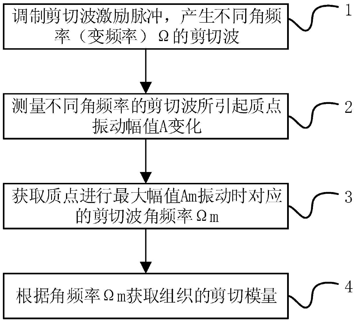
[0033] figure 1 It is a method flowchart of a preferred embodiment of the present invention. The method proposed by the present i...


PUM


Abstract

The invention discloses a multifrequency shear wave amplitude analysis-based ultrasonic elasticity imaging technique and relates to the field of medical imaging and the field of man-machine interfaces. The ultrasonic elasticity imaging technique comprises the following steps: modulating a drive pulse of a shear wave to enable the shear wave to generate different angular frequency shear waves; measuring vibration amplitudes of a particle in a tissue caused by the shear wave at different frequencies; comparing the particle vibration amplitudes in the shear wave under different angular frequencies to obtain a shear wave angular frequency corresponding to the maximal particle vibration amplitude; and acquiring the wave velocity of the shear wave or a shear modulus of the tissue or the wave velocity of the shear wave and the shear modulus of the tissue according to the angular frequency. According to the ultrasonic elasticity imaging technique, the amplitude is subjected to measurement of arelative value, rather than measurement of an absolute value, and amplitude information has higher interference resistance when being measured, relative to conventional phase information in the priorart. The ultrasonic elasticity imaging technique can be implemented through two ultrasonic vibration elements, is easy to realize portability, and has a great application potential in the field of man-machine interfaces.

Description

technical field [0001] The invention relates to the field of medical imaging and the field of man-machine interface, in particular to an ultrasonic elastic imaging technology based on multi-frequency shear wave amplitude analysis. Background technique [0002] Ultrasound elastography is an emerging tissue imaging technology in recent years, which can be used to obtain the elasticity information of human tissue, and the normal tissue and diseased tissue of the human body show different elasticity, so this technology has been widely studied in the field of medical diagnosis and apply. [0003] On the other hand, the human-machine interface can collect physiological signals of the human body, and can obtain the motion intention of the human body through processing, which can be further used to control external devices. And tissues such as skeletal muscle also have different elastic coefficients under different exercise states. Therefore, ultrasound elastography can provide hu...

Claims


Application Information

Patent Timeline
Patent Type & Authority Applications(China)
IPC IPC(8): A61B8/00A61B8/08
CPCA61B8/485A61B8/08A61B8/5207A61B8/4444A61B8/4483
Inventor 刘洪海闫纪朋杨兴晨陈镇枫赵诣王志永
Owner 交浦科技(深圳)有限公司
Why Eureka
Try Eureka