An auxiliary device for detecting peripheral vascular disease

A peripheral vascular disease and auxiliary device technology, applied in the direction of organ movement/change detection, diagnostic recording/measurement, application, etc., can solve the problems of physical exertion and peripheral vascular disease for a long time, so as to reduce physical exertion and save manpower time Low cost and practical effect

Active Publication Date: 2020-11-06
HENAN PROVINCE HOSPITAL OF TCM THE SECOND AFFILIATED HOSPITAL OF HENAN UNIV OF TCM
View PDF7 Cites 0 Cited by
  • Summary
  • Abstract
  • Description
  • Claims
  • Application Information

AI Technical Summary

Problems solved by technology

[0009] In view of the above situation, in order to overcome the defects of the prior art, the present invention provides a peripheral vascular disease detection auxiliary device, which effectively solves the problem that the existing peripheral vascular disease takes a long time in the visual detection process, and the patient's limbs are lifted by themselves or Lifting in the form of doctor's help is a lot of physical exertion for patients or doctors, but there are many deficiencies in the use of medical device testing

Method used


Image

  • An auxiliary device for detecting peripheral vascular disease
  • An auxiliary device for detecting peripheral vascular disease
  • An auxiliary device for detecting peripheral vascular disease

Examples


Embodiment 1

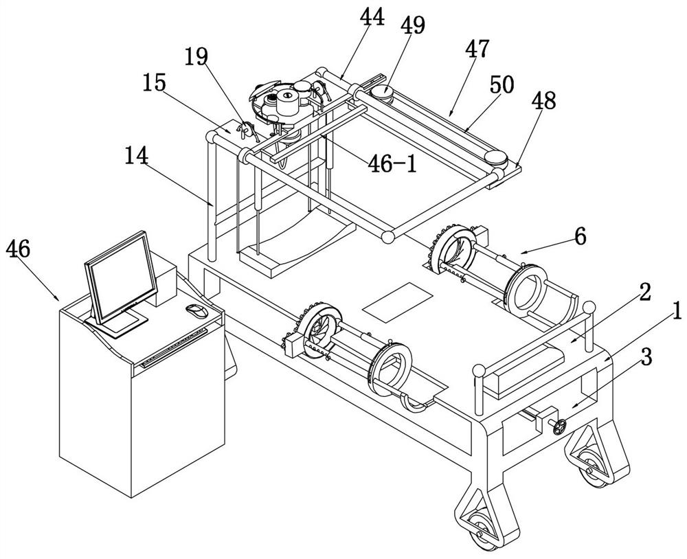
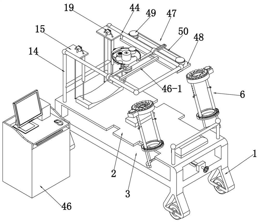
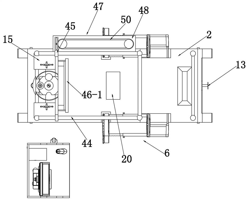
[0042] Embodiment 1, the present invention is an auxiliary device for detecting peripheral vascular disease, please refer to figure 1 , including a detection bed 1. When in use, the patient lies on the upper end of the detection bed 1 for detection. The detection bed 1 is provided with a bed board 2 and a storage board 3 placed at the lower end of the bed board 2. The lower end of the front end of the bed board 1 is along the left and right sides. Direction rotation is equipped with a rotating rod 4, and the rear ends of the two ends of the rotating rod 4 are fixed with arm lifting devices 6 placed on both sides of the bed board 2 through connecting rods 5. Please refer to Figure 7 , the rotating rod 4 and the arm lifting devices 6 on both sides are arranged in a "U" shape, and the lifting arm device 6 includes a circular disk 6-1 fixedly connected to the rotating rod 4 through a connecting rod 5, and the circular ring The rear end of the disk 6-1 is connected with the second...

Embodiment 2

[0045] Embodiment 2, on the basis of Embodiment 1, in order to make the legs lifted better, please refer to Figure 4 with Figure 5 The middle position of the bed board 2 is slidingly fitted with a buttock support block 20 along the vertical direction. It is an arc surface, and the lower end of the buttock support block 20 is movably placed on the upper end of the fixed moving block 21 at the rear end of the rack 10. The upper end of the moving block 21 is fixed with two semicircular projections 22, and the two projections The block 22 is able to hold the hip support block 20 as the rack 10 moves towards the rear end.

Embodiment 3

[0046] Embodiment 3, on the basis of Embodiment 1, in order to make this device suitable for patients of different sizes, please refer to figure 1 , figure 2 , Image 6 with Figure 9 , the rotating rod 4 is set to be telescopic, and the rotating rod 4 includes the main rotating rod 23 in the middle position and the auxiliary rotating rod 24 that is slidingly fitted on both sides. The rotating rods 24 are all set to a rectangle, and the purpose of being arranged as a rectangle is to realize the telescopic effect, and when the main rotating rod 23 rotates, it can also realize the synchronous rotation of the driving auxiliary rotating rod 24, and the auxiliary rotating rod 24 is placed on the The end of the main rotating rod 23 internal slideway is fixedly connected with the end of the main rotating rod 23 internal slideway by a spring 25;

[0047] The telescopic rod 7 includes a first telescopic rod 26 connected to the annular disk 6-1, and a second telescopic rod 27 connec...


PUM


Abstract

The invention relates to an auxiliary device for detecting peripheral vascular diseases, which effectively solves the problem that the inspection process of existing peripheral vascular disease is long, the patient's limbs are lifted up by themselves or the doctor helps to lift up, the physical output is large, and many problems are existed by using a medical device for detection; The auxiliary device comprises a detection bed, the detection bed has a bed plate and a storage board, and a lower end of the front end position of the bed plate is rotated and matched with a rotating rod, a liftingarm device is fixed at both ends of the rotating rod, the lifting arm device comprises an annular disk fixed with the rotating rod through a connecting rod, the rear end of the annular disk is connected with a second annular disk through a telescopic rod, the second annular disk is provided with a wrist stirrup device, the outer side of the rotating rod is sleeved with a gear, and the upper end ofthe storage board is slidably engaged with a rack meshing with the gear; the rack is provided with a threaded hole, the threaded hole is screwed with a screw, and the front end of the screw is fixedwith a rotating handle. The auxiliary device has the advantages of ingenious structure, strong practicability, good auxiliary inspection effect, and greatly improves the working efficiency of the doctor.

Description

technical field [0001] The invention relates to the technical field of peripheral vascular disease detection, in particular to an auxiliary device for detection of peripheral vascular disease. Background technique [0002] Clinically, vascular diseases other than cardiovascular and cerebrovascular diseases are collectively referred to as peripheral vascular diseases, including diseases of the three systems of arteries, veins and lymphatic systems; according to the World Health Organization survey, peripheral vascular diseases are a series of highly harmful high-incidence species. If it does not heal for a long time, the disease will develop progressively, and severe cases will lead to amputation, disability, and even life-threatening. [0003] Routine inspection methods for such diseases include: visual inspection, color Doppler ultrasonography, angiography, CT and MRI inspection, skin temperature tester, infrared thermal imaging detection, etc.; [0004] The visual inspect...

Claims


Application Information

Patent Timeline
Patent Type & Authority Patents(China)
IPC IPC(8): A61G13/00A61G13/10A61G13/12A61B5/00A61B8/00A61B8/08
Inventor 袁向科江瑞江泽
Owner HENAN PROVINCE HOSPITAL OF TCM THE SECOND AFFILIATED HOSPITAL OF HENAN UNIV OF TCM
Why Eureka
Try Eureka