
A method for locating fovea based on color retinal fundus image of lesion focus

A fundus image and positioning method technology, which is applied in image enhancement, image analysis, image data processing, etc., can solve the problems of large horizontal ridge noise interference, complex calculation, and difficulty in fitting a parabolic model, etc., to achieve accurate positioning, The effect of avoiding interference

Active Publication Date: 2019-03-08
UNIV OF SHANGHAI FOR SCI & TECH
View PDF8 Cites 3 Cited by
  • Summary
  • Abstract
  • Description
  • Claims
  • Application Information

AI Technical Summary

Problems solved by technology

This method of parabolic fitting of blood vessels relies on the precise detection and positioning of blood vessels and optic discs, and it is difficult to fit an accurate parabolic model in images with lesions
At the same time, the parabolic model involves the transformation between two coordinate systems, the calculation is relatively complicated, and the horizontal ridge represented by the main axis is greatly disturbed by noise

Method used


Image

  • A method for locating fovea based on color retinal fundus image of lesion focus
  • A method for locating fovea based on color retinal fundus image of lesion focus
  • A method for locating fovea based on color retinal fundus image of lesion focus

Examples


Embodiment Construction

[0034] The fovea positioning method based on the focal color retinal fundus image of the present invention will be described in more detail below in conjunction with the schematic diagram, wherein a preferred embodiment of the present invention is shown, it should be understood that those skilled in the art can modify the present invention described here, and The advantageous effects of the invention are still achieved. Therefore, the following description should be understood as the broad knowledge of those skilled in the art, but not as a limitation of the present invention.

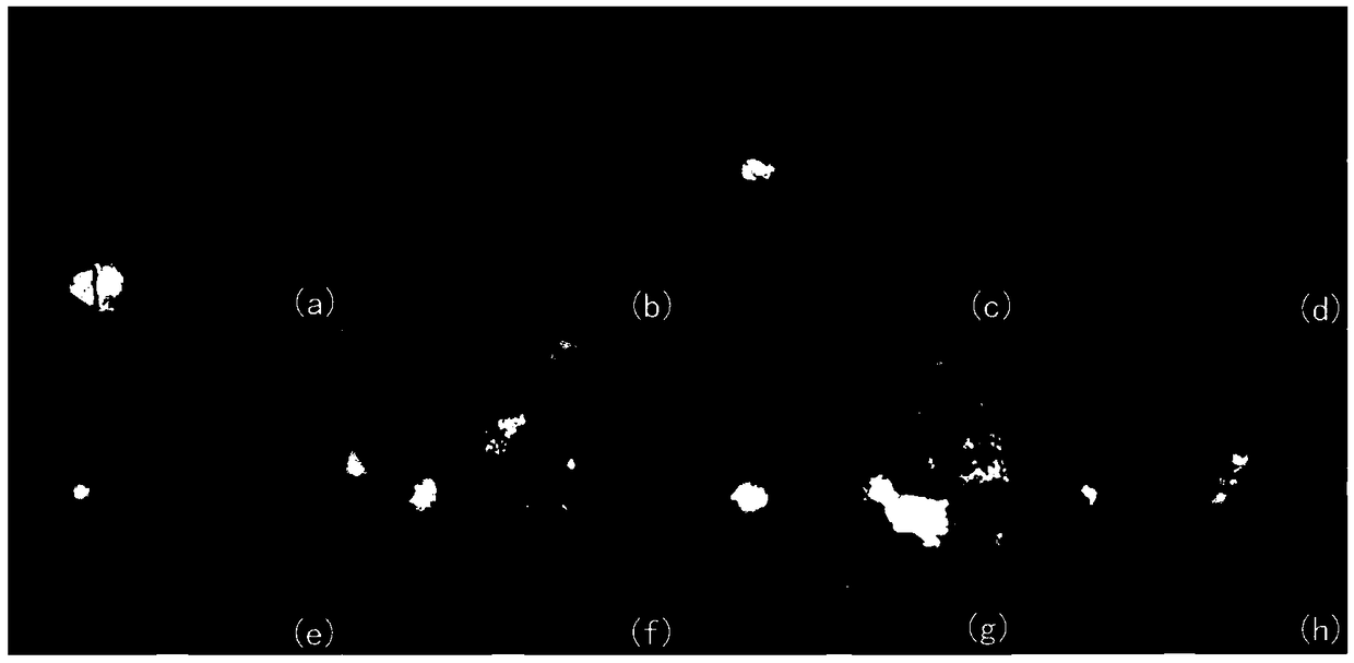
[0035] figure 1 These are 8 images from the Kaggle dataset, all of which have lesions or trauma and complex internal lighting. (a)-(d) has serious local uneven illumination; (e) contains the trauma left by laser surgery, and (f)-(h) contains large lesions, and these lesions or traumas appear in Near the macula, there is greater interference with the positioning of the fovea.

[0036] Such as figur...


PUM


Abstract

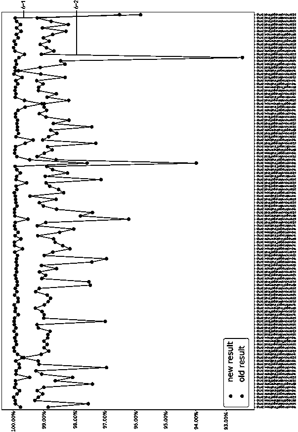
A method for locating fovea based on color retinal fundus image of lesion focus includes detecting and locating optic disc and blood vessel, acquiring blood vessel vector and blood vessel vector set,estimating horizontal ridge line passing through central fossa from blood vessel vector set, acquiring search area according to optic disc and horizontal ridge line, and acquiring the central fossa insearch area. The method of the invention determines the horizontal ridge line of the fundus image by constructing a blood vessel vector set model and calculating the vector sum of the vectors, determines a search area by utilizing the position relationship between the optic disc and the central fossa, and finally determines the central fossa positioning according to the local brightness extreme value of the search area. The method can effectively avoid the interference of the focus and illumination on the optic disc, and accurately locate the fovea.

Description

technical field [0001] The invention belongs to the technical field of retinal fundus image detection, and in particular relates to a fovea positioning method based on color retinal fundus images of lesions. Background technique [0002] The macula is one of the anatomical structures of the retinal fundus image. In normal color retinal fundus images, the macula is an area that is darker than surrounding tissue. The macula appears darker on fundus images due to the higher proportion of carotenoids present in this area. The center of the macula, called the fovea, is the darkest spot in the entire macular area. The macula is different from other anatomical structures such as blood vessels and optic discs, which have obvious structural features, while the macula and fovea only have brightness features. In brightness characteristics, the macula is very similar to dark lesions such as hemorrhages. When the fundus image shows dark lesions, it is generally difficult to accuratel...

Claims


Application Information

Patent Timeline
IPC IPC(8): G06T7/00G06T7/64
CPCG06T7/0012G06T2207/20061G06T2207/20081G06T2207/30041G06T2207/30101G06T7/64
Inventor 傅迎华李江
Owner UNIV OF SHANGHAI FOR SCI & TECH
Learn More