
Kit for detecting CYP3A4 and CYP3A5 polymorphic sites and method thereof

A CYP3A4, polymorphic site technology, applied in biochemical equipment and methods, DNA / RNA fragments, recombinant DNA technology, etc., can solve the problem of inaccurate detection of gene polymorphisms, low sensitivity and specificity, Long detection time and other problems, to achieve the effect of fast detection speed, simple result analysis and short reaction time

Active Publication Date: 2018-08-17
浙江鼎创医疗科技有限公司
View PDF5 Cites 9 Cited by
  • Summary
  • Abstract
  • Description
  • Claims
  • Application Information

AI Technical Summary

Problems solved by technology

The sensitivity and specificity of these two methods are not high, and they cannot accurately detect gene polymorphisms
In addition, most direct sequencing uses outsourced experiments, and the detection time is too long, at least 24 hours, which brings inconvenience to the detection

Method used


Image

  • Kit for detecting CYP3A4 and CYP3A5 polymorphic sites and method thereof
  • Kit for detecting CYP3A4 and CYP3A5 polymorphic sites and method thereof
  • Kit for detecting CYP3A4 and CYP3A5 polymorphic sites and method thereof

Examples


Embodiment 1

[0075] Example 1: Collection of samples

[0076] Thirty minutes before the sample collection, the patient rinsed his mouth with clean water, and should not eat or drink within 30 minutes.

[0077] 1. Open the oral swab package, carefully take out the swab, and rub the swab head against the inner wall of the oral cavity for 20-30 times.

[0078] 2. Open the saliva storage tube, break the swab head in the storage tube, cover the storage tube cap, and complete the sample collection.

Embodiment 2

[0079] Embodiment 2: DNA extraction

[0080] In this extraction step, the saliva DNA extraction kit (magnetic bead method) of Mohe Medical Technology (Shanghai) Co., Ltd. was used for extraction. Samples 1, 2, and 3 to be tested are extracted.

[0081] 1. The collected samples are placed on a constant temperature oscillating mixer, and the parameters are selected at 56°C, 15min, and 800rpm / min. Perform saliva sample pretreatment.

[0082] 2. Open the kit, take out the tube, shake it up and down 6-10 times to mix the magnetic beads in hole ⑦ of the tube, and carefully tear off the sealing film;

[0083] 3. After adding 300 μL of processed saliva sample to well ①, add 100 μL of elution buffer to well ⑨, place the tube in the drawer of the CTC-2000 nucleic acid extractor, and install the magnetic rod cover. Select the saliva DNA program to run.

[0084] 4. After the program is over, collect the extracted DNA in well ⑨ for subsequent experiments or store at -20°C.

[0085] Af...

Embodiment 3

[0086] Example 3 Fluorescent PCR detection

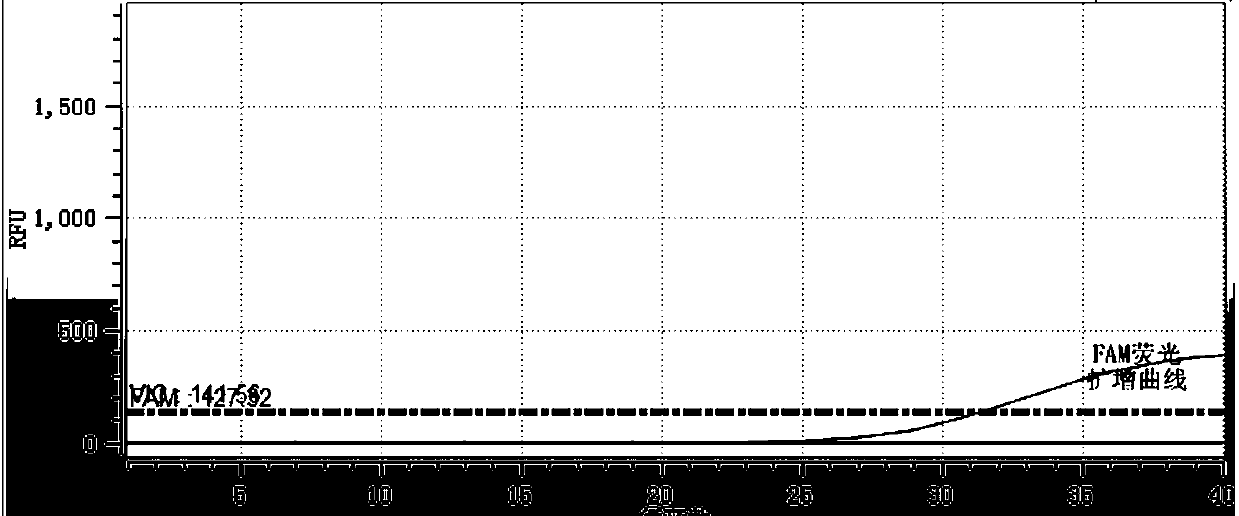
[0087] 1. Sequence 1 and sequence 2 of CYP3A4*1G specific upstream and downstream primers, specific TaqMan-MGB dual fluorescent probe sequence 5 and sequence 6; CYP3A4*1G specific upstream and downstream primers sequence 3 and sequence 4, specific TaqMan- MGB dual fluorescent probe sequence 7 and sequence 8; CYP3A5*3 specific upstream and downstream primer sequence 9 and sequence 10, specific TaqMan-MGB dual fluorescent probe sequence 13 and sequence 14; CYP3A5*3 specific upstream and downstream primer sequence 11 and sequence 12, specific TaqMan-MGB dual fluorescent probe sequence 15 and sequence 16 were divided into two groups (group A and group B respectively) for PCR experiments, as shown in the following table:

[0088]

[0089] 2. Prepare a fluorescent quantitative PCR reaction system: including PCR amplification reagents, upstream and downstream specific primers, specific wild-type TaqMan fluorescent probes, specific mutan...


PUM


Abstract

The invention discloses a kit for detecting CYP3A4 and CYP3A5 polymorphic sites and a method thereof. The kit comprises specificity upstream and downstream primer sequences of CYP3A4*1G shown in sequences 1-4 in a sequence table, specificity TaqMan-MGB bi-fluorescence probe sequences of CYP3A4*1G shown in sequences 5-8, specificity upstream and downstream primer sequences of CYP3A5*3 shown in sequences 9-12 and specificity TaqMan-MGB bi-fluorescence probe sequences of CYP3A5*3 shown in sequences 13-16. The kit has the advantages that the specificity is high, pollution is prevented, the sensitivity is high, the detection speed is high, result analysis is simple, and the accuracy is high; the kit can judge the metabolism capacity of to-be-detected objects to fentanyl medicine by detecting polymorphic sites of CYP3A4 and CYP3A5 genes, the using amount of medicine such as fentanyl can be more accurately and effectively guided accordingly, and the kit has the actual clinical application value. The kit is applied to clinical diagnosis, the fit degree between the kit and the prior art is higher, the cost is low, and repeatability is high.

Description

technical field [0001] The invention relates to the field of biotechnology, in particular to gene detection, more specifically to a CYP3A4, CYP3A5 polymorphism site gene detection kit and a method thereof. Background technique [0002] CYP3A4 is the most important member of the CYP3A subfamily, accounting for 30%-40% of the total P450 enzymes, mainly distributed in the liver and intestinal tract. It participates in the oxidative metabolism of many drugs and exogenous toxicants, and can metabolize about 50% of commonly used clinical drugs. Studies have shown that there are obvious individual differences in the expression and activity of CYP3A4 in human liver, and 90% of these differences are related to genetic polymorphisms. The CYP3A4 gene is located on human chromosome 7 7q21.1-q22.1, about 27 kbp in length, and its gene structure includes 13 exons and 12 introns. CYP3A4*1G (20230G>A) is a single nucleotide polymorphism (SNP) in intron 10 of CYP3A4, and its frequency i...

Claims


Application Information

Patent Timeline
Patent Type & Authority Applications(China)
IPC IPC(8): C12Q1/6858C12N15/11
CPCC12Q1/6858C12Q2561/101C12Q2563/107C12Q2527/125
Inventor 唐荣苏丽吴丹
Owner 浙江鼎创医疗科技有限公司
Learn More