Nano-CT 3D Imaging Method with Three Quantitative Imaging Mechanisms

A three-dimensional imaging and mechanism technology, applied in the direction of measuring devices, instruments, scientific instruments, etc.

Active Publication Date: 2019-08-13
济南汉江光电科技有限公司
View PDF4 Cites 0 Cited by
  • Summary
  • Abstract
  • Description
  • Claims
  • Application Information

AI Technical Summary

Problems solved by technology

[0006] The object of the present invention is to provide a nano-CT three-dimensional imaging method with three quantitative imaging mechanisms to solve the problems encountered in the quantification of the depth of field and phase contrast of the existing nano-CT. Resolution 3D Imaging Provides Solutions

Method used


Image

  • Nano-CT 3D Imaging Method with Three Quantitative Imaging Mechanisms
  • Nano-CT 3D Imaging Method with Three Quantitative Imaging Mechanisms
  • Nano-CT 3D Imaging Method with Three Quantitative Imaging Mechanisms

Examples


Embodiment Construction

[0029] An embodiment of the present invention provides a nano-CT three-dimensional imaging method with three quantitative imaging mechanisms, comprising the following steps:

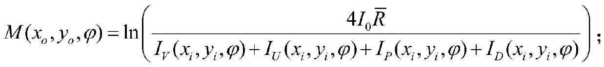
[0030] Through three quantitative imaging mechanisms, the two-dimensional absorption contrast image, two-dimensional refraction contrast image and two-dimensional scattering contrast image of each corner of the sample are reconstructed on the object surface of the X-ray differential phase contrast microscope. Among them,

[0031] The expression of the two-dimensional absorption contrast image reconstructed at each corner of the sample on the object surface of the X-ray differential phase contrast microscope is:

[0032]

[0033]The expression for reconstructing the two-dimensional refraction contrast image of each corner of the sample on the object surface of the X-ray differential phase contrast microscope is:

[0034]

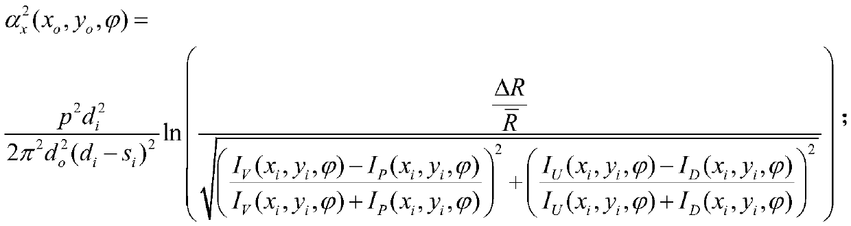
[0035] The expression for reconstructing the two-dimensional scattering contr...


PUM

PropertyMeasurementUnit
diameteraaaaaaaaaa
sizeaaaaaaaaaa
diameteraaaaaaaaaa

Abstract

The invention discloses a nanometer CT three-dimensional imaging method using three quantitative imaging mechanisms. The method comprises taking a valley macrophotograph, an upslope macrophotograph, apeak macrophotograph and a downslope macrophotograph of a sample at different angles on the image plane of an X-ray differential phase contrast microscope according to an angle signal response imaging mechanism, rebuilding a two-dimensional absorption contrast image, a two-dimensional refraction contrast image and a two-dimensional scattering contrast image of the sample at different angles on the object plane of the X-ray differential phase contrast microscope according to three quantitative imaging mechanisms, and rebuilding a linear absorption coefficient three-dimensional image, a linearscattering coefficient three-dimensional image, a refractive index real attenuation rate three-dimensional image and a refractive index real attenuation rate derivative three-dimensional image of thesample according to inverse Radon conversion. The method can realize three-dimensional imaging of whole cells with diameters of 5 micrometers or more and has the nanometer scale resolution rate.

Description

technical field [0001] The invention relates to the technical field of nano-resolution X-ray microscope imaging, in particular to a nano-CT three-dimensional imaging method with three quantitative imaging mechanisms. Background technique [0002] Nano-CT with X-ray microscope as the core is another crystallization of the development of human science and technology after optical microscope and electron microscope. It is opening a new window for people to observe the microscopic world and has become a new growth point for the development of human science. Like visible light, X-rays have various imaging mechanisms and rich sources of contrast. Information such as absorption, phase shift, refraction, scattering, and fluorescence can be used to observe and analyze various physical changes, chemical reactions, and nanostructures. [0003] X-ray nano-CT technology has played a powerful role in promoting the development of many disciplines. However, it is still far from meeting the ...

Claims


Application Information

Patent Timeline
Patent Type & Authority Patents(China)
IPC IPC(8): G01N23/046
CPCG01N23/046
Inventor 朱佩平何其利廖可梁袁清习张凯黄万霞朱中柱王研
Owner 济南汉江光电科技有限公司
Why Eureka
Try Eureka