Novel growth/neurotrophic factor composition for inducing mature neuron division in vivo and purpose thereof
A technology of nerve growth factor and growth factor, which is applied in the direction of drug combination, nervous system diseases, organic active ingredients, etc., and can solve problems such as low enough to compensate neurons
- Summary
- Abstract
- Description
- Claims
- Application Information
AI Technical Summary
Problems solved by technology
Method used
Image
Examples
Embodiment 1
[0062] Example 1: Induction of dividing neurons in the adult rat cerebral cortex
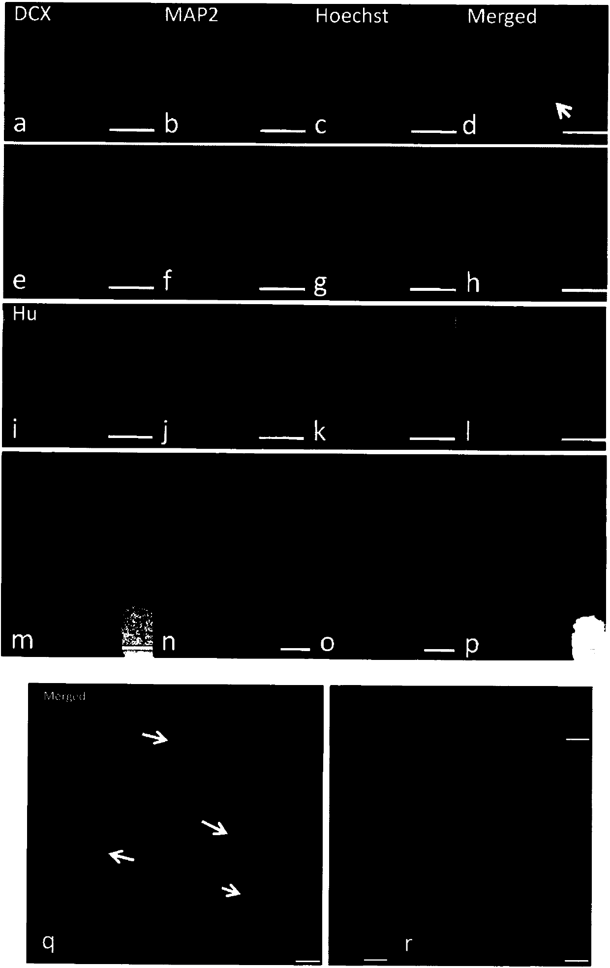
[0063] The present inventors combined (n=3) or did not contain (n=3) fibrin (fibrin) matrix (12.5IU / ml fibrinogen and 12.5IU / ml plasminogen, Sigma F6755 and T5772) of the present invention Compositions (growth factor / neurotrophic factor combination + T3) were injected into the cerebral cortex of adult rats respectively. Wherein, the growth factor / neurotrophic factor combination is composed of the following: EGF (Sigma E4127Lot: SLBJ4118V), bFGF (Invitrogen PMG0033, Lot 489821E), HGF (Millipore Cat: 375228-5UG, Lot: D00165582), IGF (Millipore GF306Lot :2576396), NGF (extracted from mouse submandibular gland, kindly provided by Prof.Shao N) and BDNF. There was no difference in the number of dividing neurons observed between the two groups. Therefore, the growth factor / neurotrophic factor combination + T3 was chosen for subsequent experiments.
[0064] The inventors observed that by microdialysi...
Embodiment 2
[0070] Example 2: Spinal Cord Projection
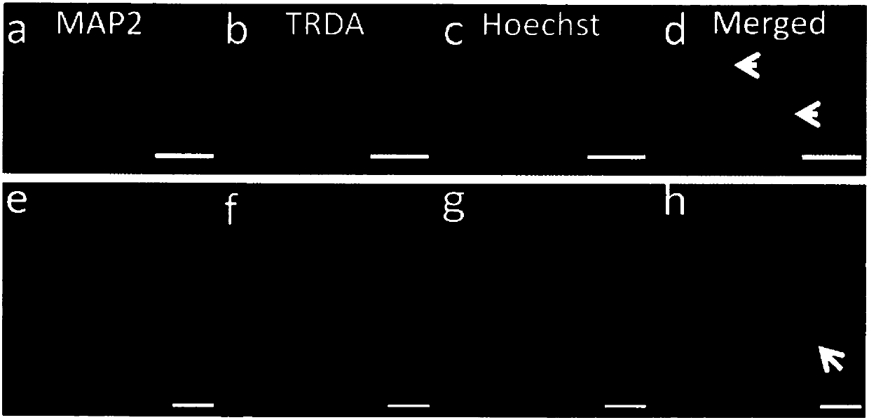
[0071] In order to identify whether the split-induced neurons in the V layer of the cerebral cortex project to the spinal cord, the inventors injected 3kDa Texas red-dextran amine (TRDA) into the cortex on both sides of the C5 segment in advance. in the spinal cord. In the first week after TRDA injection, dense TRDA-tracked nerve fibers in the C3 spinal cord tract were observed. However, with the prolongation of survival time, the fluorescence intensity gradually became lower and was not observed 14 days after retrograde tracing. This suggests that at the injection site, the regenerated fibers (if any) do not absorb TRDA.
[0072] Then, the composition of the present invention was injected into the M1 region of the cerebral cortex of rats that had received TRDA injection for 4 or 8 weeks, and then the animals were maintained for another 2 days before labeling brain and spinal cord sections with Hoechst and anti-MAP2 antibodies. suc...
Embodiment 3
[0074] Example 3: Inducing the fate of dividing neurons
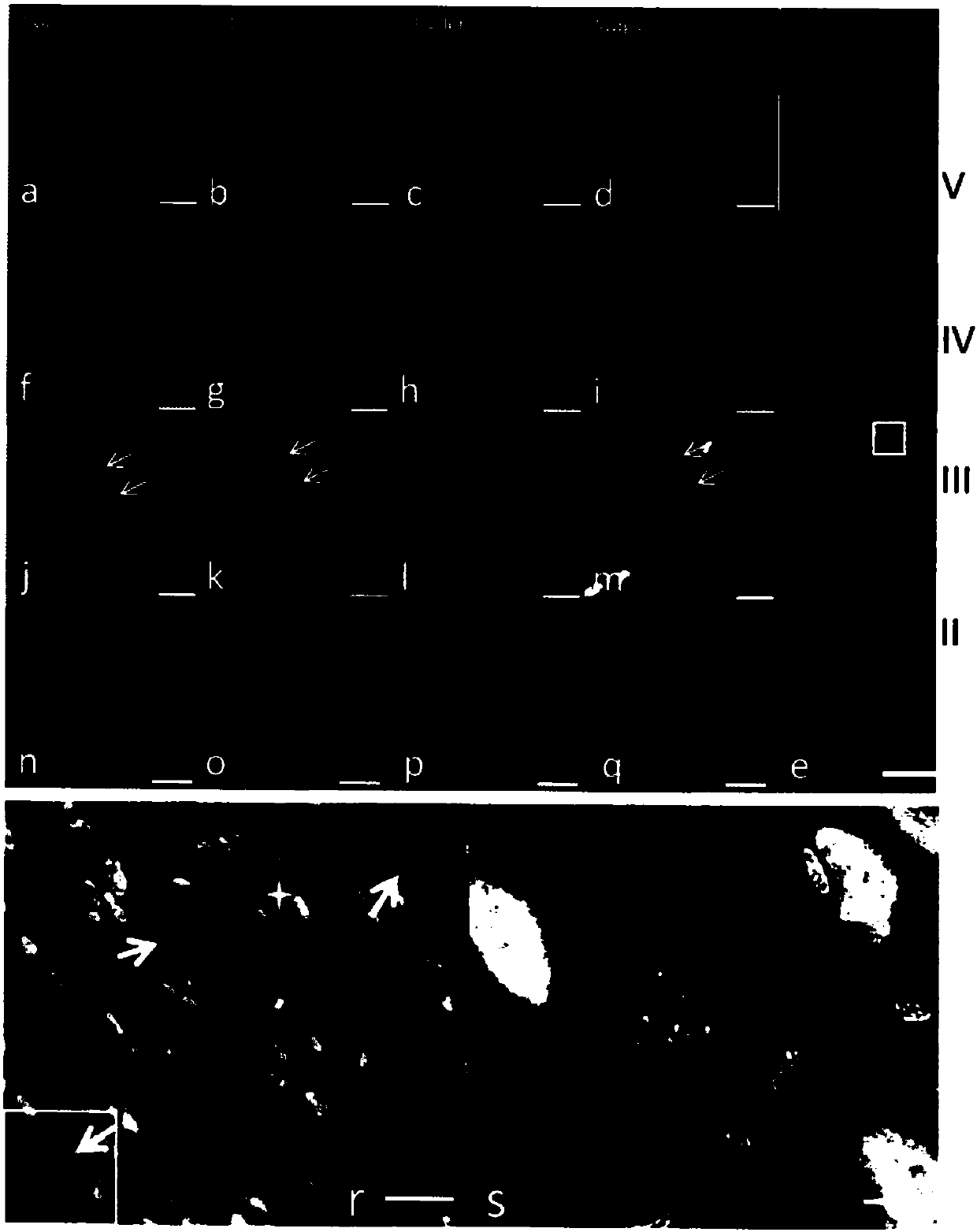
[0075] To examine the survival of post-mitotic neurons, the inventors injected the growth factor / neurotrophic factor combination + T3 into the cerebral cortex M1 together with BrdU intraperitoneally, administered 3 times within 36 hours to avoid triggering endogenous neural precursors cell. Animals were continued for 4 or 8 weeks. The results showed that all BrdU + / NeuN + Neurons were distributed within a 2 mm diameter of the cylindrical area around the injection needle in the cerebral cortex. Compared to animals sacrificed immediately after induction, in BrdU + NeuN expression was significantly restored in neurons (see Figure 4 ). From the morphology point of view, BrdU + / NeuN + Neurons can be divided into four types: multipolar neurons ( Figure 4 a-d), bipolar neurons ( Figure 4 e-h), pyramidal cells ( Figure 4 i-l) and large neurons ( Figure 4 m-s).
[0076] In the 4-week group, BrdU + / NeuN + T...
PUM
 Abstract
Description
Claims
Application Information
 - R&D
- Intellectual Property
- Life Sciences
- Materials
- Tech Scout
- Unparalleled Data Quality
- Higher Quality Content
- 60% Fewer Hallucinations
Browse by: Latest US Patents, China's latest patents, Technical Efficacy Thesaurus, Application Domain, Technology Topic, Popular Technical Reports.
© 2025 PatSnap. All rights reserved.Legal|Privacy policy|Modern Slavery Act Transparency Statement|Sitemap|About US| Contact US: help@patsnap.com


