Method and system for automated brain tumor diagnosis using image classification

An image-in-image technology, applied in the field of automatic brain tumor diagnosis, which can solve problems such as pathologist burden

Inactive Publication Date: 2018-01-02
SIEMENS AG
View PDF4 Cites 9 Cited by
  • Summary
  • Abstract
  • Description
  • Claims
  • Application Information

AI Technical Summary

Problems solved by technology

However, manual judgment for diagnosis can be subjective and variable for different pathologists
In addition, optical biopsy-based diagnostic tasks can be a significant burden for pathologists due to the large amount of image data acquired

Method used


Image

  • Method and system for automated brain tumor diagnosis using image classification
  • Method and system for automated brain tumor diagnosis using image classification
  • Method and system for automated brain tumor diagnosis using image classification

Examples


Embodiment Construction

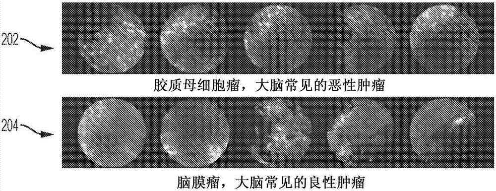
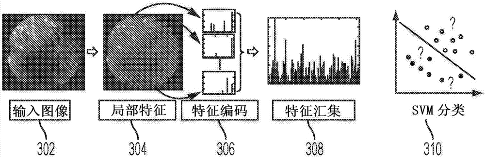
[0014] The present invention relates to automatic classification of different types of tissue in medical images using machine learning based image classification. Embodiments of the present invention may be applied to endoscopic images of brain tumor tissue for automated brain tumor diagnosis. Embodiments of the present invention are described herein to visually understand an automatic classification method of tissue in medical images. Digital images often consist of digital representations of one or more objects (or shapes). Digital representations of objects are generally described herein in terms of recognizing and manipulating objects. Such manipulations are virtual manipulations implemented in memory or other circuitry / hardware of a computer system. Accordingly, it should be understood that embodiments of the present invention may be implemented within a computer system using data stored within the computer system.

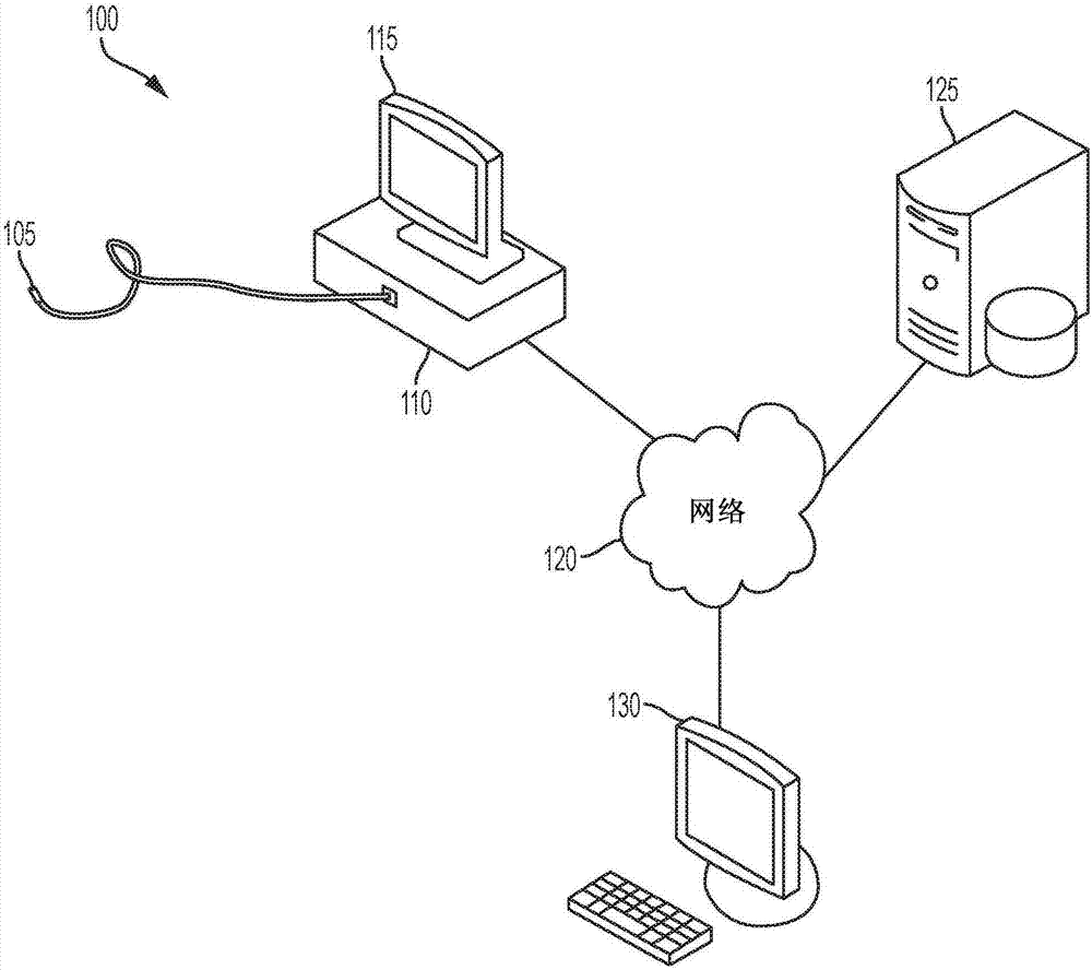
[0015] figure 1 An example of a system 100 for acqu...


PUM


Abstract

A method and system for classifying tissue endomicroscopy images are disclosed. Local feature descriptors are extracted from an endomicroscopy image. Each of the local feature descriptors is encoded using a learnt discriminative dictionary. The learnt discriminative dictionary includes class-specific sub-dictionaries and penalizes correlation between bases of sub-dictionaries associated with different classes. Tissue in the endomicroscopy image is classified using a trained machine learning based classifier based on the coded local feature descriptors encoded using a learnt discriminative dictionary.

Description

[0001] This application claims the benefit of US Provisional Application 62 / 139,016, filed March 27, 2015, the disclosure of which is incorporated herein by reference. technical field [0002] The present invention relates to classifying different types of tissue in medical image data using machine learning based image classification, and more particularly to automated brain tumor diagnosis using machine learning based image classification. Background technique [0003] Cancer is a major health problem around the world. Early diagnosis of cancer is critical to the success of cancer treatment. Traditionally, pathologists obtain histopathological images of biopsies sampled from patients, examine the histopathological images under a microscope, and make judgments about the diagnosis based on their knowledge and experience. Unfortunately, intraoperative rapid histopathology is often insufficient to provide pathologists with accurate diagnostic information. Biopsies are often n...

Claims


Application Information

Patent Timeline
Patent Type & Authority Applications(China)
IPC IPC(8): G06K9/62G06K9/00G06V10/772
CPCG06V20/698G06V10/772A61B1/000094G06F18/28A61B5/4255G06T7/0012
Inventor 万韶华孙善辉苏巴布拉塔·巴塔查里亚陈德仁阿里·卡门
Owner SIEMENS AG
Why Eureka
Try Eureka