Ultrasonic peeping probe and elastic imaging system and method
A technology of elastography and ultrasound, applied in the direction of ultrasound/sonic/infrasonic Permian technology, ultrasound/sonic/infrasonic image/data processing, ultrasound/sonic/infrasonic diagnosis, etc. It can solve the problems of applying pressure and achieve operation Effects of simplicity, pain reduction, and system cost reduction
- Summary
- Abstract
- Description
- Claims
- Application Information
AI Technical Summary
Problems solved by technology
Method used
Image

Examples
Embodiment 1
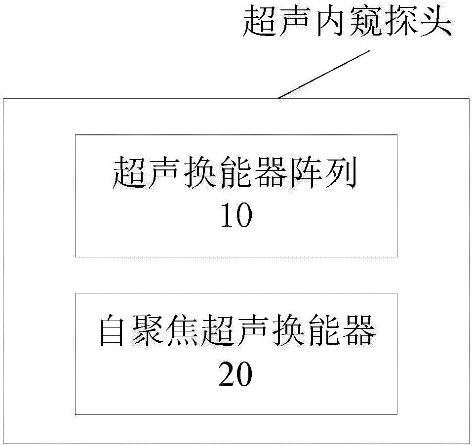
[0031] figure 1 It shows a schematic structural diagram of an ultrasound endoscopic probe according to an embodiment of the present invention, which is used to obtain ultrasound echo signals before and after the biological tissue is compressed when the biological tissue is elastically imaged. It should be supplemented that the biological tissues here include blood vessels, gastrointestinal tissues, etc., as well as tissues in organs.
[0032] according to figure 1 As shown, the ultrasound endoscopic probe includes an ultrasound transducer array 10 and a self-focusing ultrasound transducer 20. The ultrasonic transducer array 10 includes a plurality of ultrasonic transducers for transmitting ultrasonic waves and receiving ultrasonic echoes reflected by the biological tissues to be detected. The self-focusing ultrasound transducer 20 is used to generate a focused ultrasound beam, which will generate acoustic radiation force to act on the biological tissue to be tested, thereby causi...
Embodiment 2
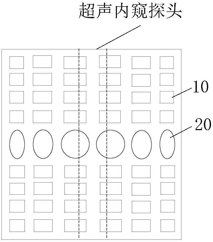
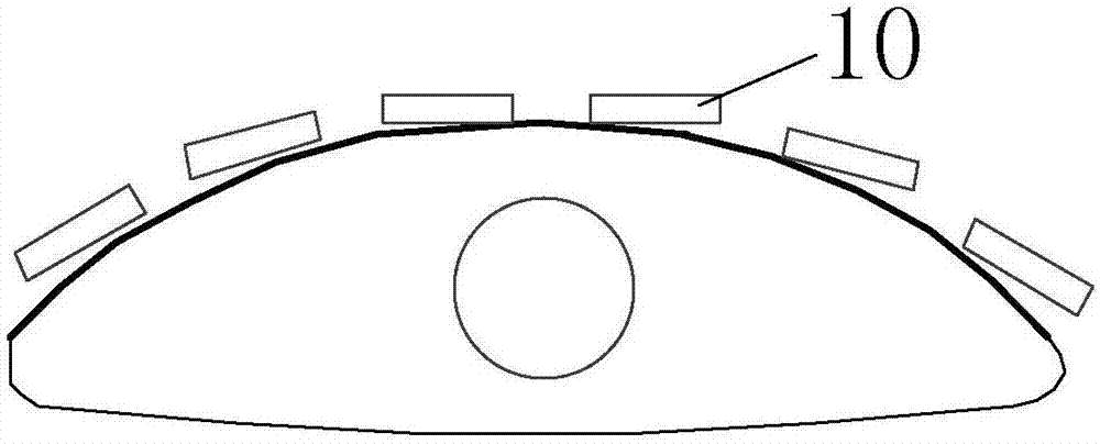
[0035] figure 2 , image 3 with Figure 4 A top view, a front view, and a side view of an ultrasound endoscopic probe according to an embodiment of the present invention are respectively shown. The ultrasound endoscopic probe is used to obtain the compression before and after the biological tissue is subjected to elastic imaging. Ultrasonic echo signal. It should be supplemented that the biological tissues here include blood vessels, intestines and stomach, etc., and can also be tissues in organs.
[0036] Such as figure 2 As shown, the ultrasound endoscopic probe includes an ultrasound transducer array 10 and a self-focusing ultrasound transducer 20. Please refer to the first embodiment for a specific description of the ultrasonic transducer array 10 and the self-focusing ultrasonic transducer 20.
[0037] Such as image 3 As shown, the ultrasonic transducer array 10 forms an arc-shaped curved surface structure. Compared with the ultrasonic transducer array 10 forming a planar...
Embodiment 3
[0044] Figure 5 with Image 6 The general structure diagram and the axial cross-sectional view of another ultrasound endoscopic probe according to an embodiment of the present invention are respectively shown. The ultrasound endoscopic probe is used to obtain ultrasound before and after the biological tissue is compressed when the biological tissue is elastically imaged. Echo signal. It should be supplemented that the biological tissues here include blood vessels, intestines and stomach, etc., and can also be tissues in organs.
[0045] according to Figure 5 As shown, the ultrasound endoscopic probe includes an ultrasound transducer array 10 and a self-focusing ultrasound transducer 20. Please refer to the first embodiment for details.
[0046] Such as Figure 5 As shown, the curved surface formed by the ultrasonic transducer array 10 and the self-focusing ultrasonic transducer 20 is drum-shaped or olive-shaped. Therefore, the ultrasonic transducer array 10 forms an arc-shaped c...
PUM
 Abstract
Description
Claims
Application Information
 - Generate Ideas
- Intellectual Property
- Life Sciences
- Materials
- Tech Scout
- Unparalleled Data Quality
- Higher Quality Content
- 60% Fewer Hallucinations
Browse by: Latest US Patents, China's latest patents, Technical Efficacy Thesaurus, Application Domain, Technology Topic, Popular Technical Reports.
© 2025 PatSnap. All rights reserved.Legal|Privacy policy|Modern Slavery Act Transparency Statement|Sitemap|About US| Contact US: help@patsnap.com