A staining method for observing collagen fibers by polarized light microscope
A dyeing method and collagen fiber technology, which is applied in the field of dyeing collagen fibers observed by a polarized light microscope, can solve the problems of unclear structure of collagen fibers, cumbersome steps, weak coloring of collagen fibers, etc.
- Summary
- Abstract
- Description
- Claims
- Application Information
AI Technical Summary
Problems solved by technology
Method used
Image
Examples
Embodiment 1
[0034] Tissues were obtained from 42-day-old normal and chronic fibrotic mouse livers and fixed with 10% formaldehyde.
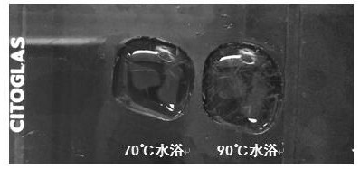
[0035] Picric acid-Sirius red dyeing solution: add 0.1g of picric acid to 1mL Sirius red dyeing solution, after a 90°C water bath, there is insoluble powder at the bottom of the dyeing solution, take the upper solution for later use.
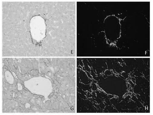
[0036]Staining method: (1) fix the tissue in 10% formaldehyde solution, routinely dehydrate and embed in wax for pathology; (2) paraffin sections with a thickness of 4 μm, routinely dewax to water to obtain tissue sections; (3) drip on the tissue sections Add picric acid-Sirius red staining solution in a water bath at 90°C for 30 minutes; (4) Add 1% w / v acetic acid aqueous solution dropwise and soak for 2 times, each time for 30 seconds; (5) Wash slightly in running water to remove the surface staining (6) Dry in an oven, and fix with neutral resin; (7) Observe under ordinary light microscope and polarized light microscope.
Embodiment 2
[0038] Tissues were obtained from 42-day-old normal and chronic fibrotic mouse livers and fixed with 10% formaldehyde.
[0039] Picric acid-Sirius red dyeing solution: add 0.1g of picric acid to 1mL Sirius red dyeing solution, after a 90°C water bath, there is insoluble powder at the bottom of the dyeing solution, take the upper solution for later use.
[0040] Staining method: (1) fix the tissue in 10% formaldehyde solution, routinely dehydrate and embed in wax for pathology; (2) paraffin sections with a thickness of 4 μm, routinely dewax to water to obtain tissue sections; (3) drip on the tissue sections Add picric acid-Sirius red staining solution in a 70°C water bath for 30 minutes; (4) Add dropwise 1% w / v acetic acid aqueous solution for 2 times, 30s each time; (5) Slightly soak in running water to remove the surface staining of tissue sections (6) Dry in an oven, and fix with neutral resin; (7) Observe under ordinary light microscope and polarized light microscope.
Embodiment 3
[0042] Tissues were obtained from 42-day-old normal and chronic fibrotic mouse livers and fixed with 10% formaldehyde.
[0043] Picric acid-Sirius red dyeing solution: add 0.1g of picric acid to 1ml Sirius red dyeing solution, after 90℃ water bath, there is insoluble powder at the bottom of the dyeing solution, take the upper solution for later use.
[0044] Staining methods: (1) Fix the tissue in 10% formaldehyde solution, routinely dehydrate and make transparent, and embed in wax; (2) Paraffin sections with a thickness of 4 μm are routinely dewaxed to water to obtain tissue sections; (3) Hematoxylin staining for 6 minutes , rinse the tissue sections slightly with running water; (4) stain the tissue sections with picric acid-Sirius red staining solution in a 90°C water bath for 30 min; (5) add 1% w / v acetic acid aqueous solution to soak twice, each time 30s; (6) Slightly immerse in running water to remove the staining solution on the surface of the tissue section; (7) Dry in ...
PUM
 Abstract
Description
Claims
Application Information
 - R&D
- Intellectual Property
- Life Sciences
- Materials
- Tech Scout
- Unparalleled Data Quality
- Higher Quality Content
- 60% Fewer Hallucinations
Browse by: Latest US Patents, China's latest patents, Technical Efficacy Thesaurus, Application Domain, Technology Topic, Popular Technical Reports.
© 2025 PatSnap. All rights reserved.Legal|Privacy policy|Modern Slavery Act Transparency Statement|Sitemap|About US| Contact US: help@patsnap.com


