
Fully convolutional network based brain MRI tumor segmentation method

A fully convolutional network and brain technology, applied in the field of medical image processing, can solve problems such as easy overfitting and difficult training of deep networks

Active Publication Date: 2017-08-04
ZHEJIANG NORMAL UNIVERSITY
View PDF5 Cites 64 Cited by
  • Summary
  • Abstract
  • Description
  • Claims
  • Application Information

AI Technical Summary

Problems solved by technology

Although the neural network has many shortcomings, a group of people such as Hinton insisted on their research and proposed many new methods to solve the problem that the deep network is difficult to train and easy to overfit.

Method used


Image

  • Fully convolutional network based brain MRI tumor segmentation method
  • Fully convolutional network based brain MRI tumor segmentation method
  • Fully convolutional network based brain MRI tumor segmentation method

Examples


Embodiment Construction

[0033] The implementation of the present invention will be described in detail below in conjunction with the accompanying drawings and examples, so as to fully understand and implement the process of how to apply technical means to solve technical problems and achieve technical effects in the present invention.

[0034] The brain MRI tumor segmentation method based on a fully convolutional network in the embodiment of the present application is used for brain MRI tumor segmentation.

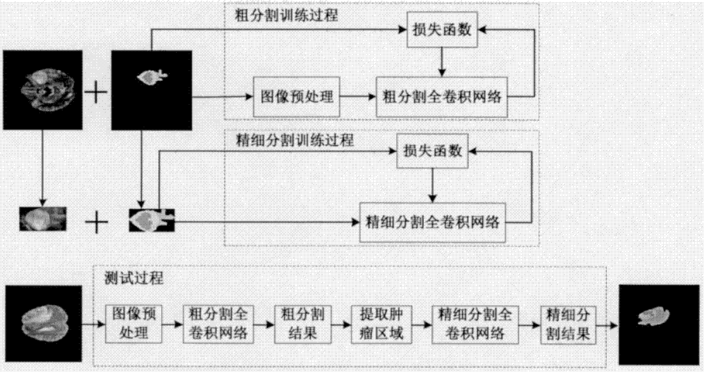
[0035] Such as figure 1 As shown, the brain MRI tumor segmentation method based on the full convolutional network of the embodiment of the present application mainly includes the following steps:

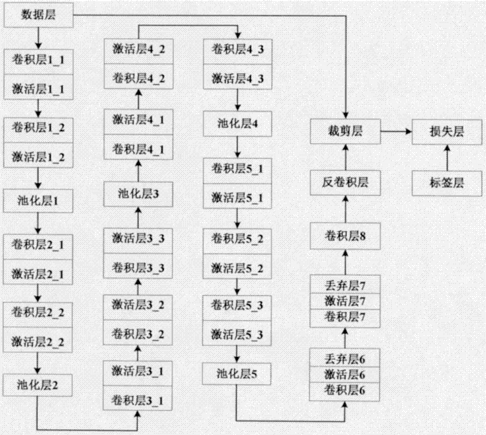
[0036] Step 1 trains a coarse segmentation fully convolutional network model for detecting tumor regions in the original image;

[0037] Step 2 trains a finely segmented fully convolutional network model for finely segmenting the internal structure of the tumor region;

[0038] Step 3 uses the traine...


PUM


Abstract

The invention discloses a fully convolutional network based tumor segmentation method in allusion to a brain MRI image. A task of tumor segmentation is divided into two steps such as coarse segmentation and fine segmentation. The fully convolutional network based brain MRI tumor segmentation method comprises the steps of firstly training a coarse segmentation fully convolutional network model by using a preprocessed sample so as to be used for detecting a tumor region in an original image, and then taking the tumor region as a training sample to train a fine segmentation fully convolutional network so as to be used for performing fine segmentation on the internal structure of the tumor region. The result shows that the method has a good segmentation effect for a brain MRI tumor.

Description

technical field [0001] The invention belongs to the technical field of medical image processing. Specifically, it relates to a brain MRI tumor segmentation method based on a fully convolutional network for the purpose of segmenting tumors in brain magnetic resonance imaging (MRI). Background technique [0002] Brain tumor is one of the tumors with the highest morbidity and mortality. MRI is a particularly effective means of evaluating brain tumors clinically. Accurate segmentation of brain tumors and tumor internal structures is not only important for assisting doctors in their treatment planning, but also for subsequent follow-up evaluations. However, the manual segmentation method is very time-consuming, and is prone to mis-segmentation due to subjective factors. Therefore, finding an accurate brain tumor segmentation method is necessary. However, due to the highly variable shape, structure, and location of the tumor area, in addition, due to factors such as imaging de...

Claims


Application Information

Patent Timeline
IPC IPC(8): G06T7/11G06N3/04G06N3/08
CPCG06T2207/10088G06T2207/20084G06T2207/20081
Inventor 张长江方明超
Owner ZHEJIANG NORMAL UNIVERSITY
Learn More