A method for establishing a liver cancer xenograft tumor model based on the hanging drop culture method of circulating tumor cells in liver cancer
A technology of biological gel and hanging drop culture, which is applied in the field of biomedicine, can solve the problems of high price and low success rate of modeling, and achieve the effect of low cost, simple preparation method and mature process
- Summary
- Abstract
- Description
- Claims
- Application Information
AI Technical Summary
Problems solved by technology
Method used
Image
Examples
Embodiment 1
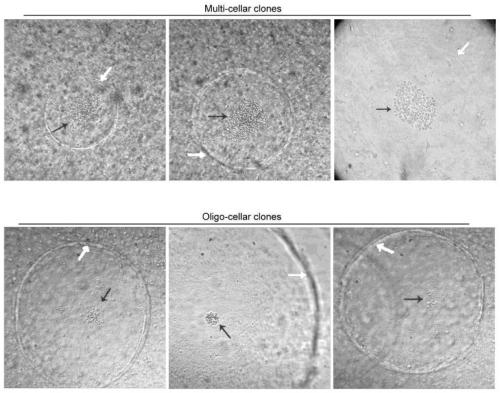
[0046] Example 1: Preoperative CTC culture results of a patient with liver cancer
[0047] 1. Experimental method:
[0048] (1) Take 10 mL of preoperative peripheral blood from the patient, mix it with HBSS buffer 1:1, and place it in a 50 mL centrifuge tube. Add 10mL of Ficoll density gradient solution to another 50mL centrifuge tube, and gently add the diluted peripheral blood to the density gradient solution to form an obvious layer. Centrifuge at room temperature at 2000rpm for 20min. At this time, the liquid is divided into 4 layers. Use a 3mL Pasteur pipette to absorb the nucleated cell layer, add 10mL of HBSS buffer, and centrifuge at 1000rpm for 5min to collect the cells and fully remove the supernatant. Add 1mL of HBSS (containing 2% serum ) to resuspend the cells and transfer to a 5 mL sterile flow tube.
[0049] (2) Add 20 μL of StemCell CD45 negative selection magnetic beads, mix gently and incubate at room temperature for 15 minutes, put them in a magnetic field...
Embodiment 2
[0056] Example 2: Detection of Subcutaneous Tumor Formation Ability of Peripheral Blood MC and OC Type Clones in Nude Mice from 7 Cases of Liver Cancer Patients
[0057] 1. Experimental method:
[0058] (1) Take 10 mL of preoperative peripheral blood from the patient, mix it with HBSS buffer 1:1, and place it in a 50 mL centrifuge tube. Add 10mL of Ficoll density gradient solution to another 50mL centrifuge tube, and gently add the diluted peripheral blood to the density gradient solution to form an obvious layer. Centrifuge at room temperature at 2000rpm for 20min. At this time, the liquid is divided into 4 layers. Use a 3mL Pasteur pipette to absorb the nucleated cell layer, add 10mL of HBSS buffer, and centrifuge at 1000rpm for 5min to collect the cells and fully remove the supernatant. Add 1mL of HBSS (containing 2% serum ) to resuspend the cells and transfer to a 5 mL sterile flow tube.
[0059] (2) Add 20 μL of StemCell CD45 negative selection magnetic beads, mix gentl...
Embodiment 3
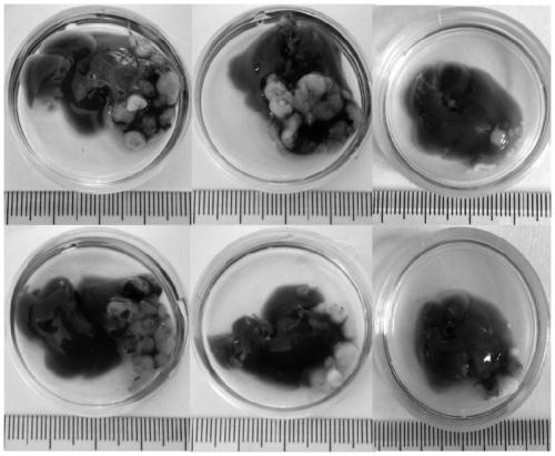
[0069] Example 3: Establishment of a PDX model of tumor formation under the liver capsule of CTC nude mice
[0070] 1. Experimental method:
[0071] (1) Pick cell clones from the CTC culture system and suspend them in 20 μL HBSS buffer for later use; after nude mice are anesthetized with isoflurane inhalation, a 0.4-0.5 cm incision is made on the upper abdomen to expose a small part of the liver, and 20 μL of the cells are injected into an insulin syringe. The suspension was injected under the liver capsule and sutured. Two weeks later, the tumor was observed by laparotomy.
[0072] (2) Embed the tumor tissue in wax block, and detect with HE staining: xylene (I) 5-10 min, xylene (II) 5-10 min, 95% ethanol (I) 1-3 min, 95% ethanol (II) 1 -3min, 80% ethanol for 1min, distilled water for 1min, hematoxylin semen staining for 5-15min, running water to wash away the hematoxylin semen for 1-3s, 1% hydrochloric acid ethanol for 1-3s, washing with water for 10-30s, and the blue-promo...
PUM
 Abstract
Description
Claims
Application Information
 - R&D
- Intellectual Property
- Life Sciences
- Materials
- Tech Scout
- Unparalleled Data Quality
- Higher Quality Content
- 60% Fewer Hallucinations
Browse by: Latest US Patents, China's latest patents, Technical Efficacy Thesaurus, Application Domain, Technology Topic, Popular Technical Reports.
© 2025 PatSnap. All rights reserved.Legal|Privacy policy|Modern Slavery Act Transparency Statement|Sitemap|About US| Contact US: help@patsnap.com


