Medical image analysis method, system and medical device

A technology of medical imaging and analysis method, applied in the field of medical imaging, can solve the problems of not being able to reflect the volume characteristics of the data, and not conducive to comparing the differences of images from different channels

Active Publication Date: 2017-07-07
SHENZHEN MINDRAY BIO MEDICAL ELECTRONICS CO LTD
View PDF8 Cites 34 Cited by
  • Summary
  • Abstract
  • Description
  • Claims
  • Application Information

AI Technical Summary

Problems solved by technology

[0004] For this reason, one of the purposes of the present invention is to provide a fast and effective way to observe the same modality for the technical problem that the display method in the prior art cannot reflect the volume characteristics of the data and is not conducive to comparing the differe...

Method used


Image

  • Medical image analysis method, system and medical device
  • Medical image analysis method, system and medical device
  • Medical image analysis method, system and medical device

Examples


Embodiment 1

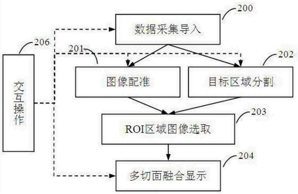
[0054] Such as figure 1 As shown, the medical image analysis method of this embodiment may include three parts: data collection and import, image analysis, and image display.

[0055] As shown in step 200, in the data collection and importing step, various types of image data can be acquired and loaded first, and then enter the image analysis part, where the image analysis can include image registration shown in step 201, image segmentation shown in step 202 , and the image selection of the region of interest shown in step 203, so as to realize the corresponding processing of each data and generate the slice image for display. Finally, as shown in step 204, the slice image sequence and parameter information generated by the image analysis part Send it to the display unit for multi-faceted fusion display.

[0056] In addition, the medical image analysis method of this embodiment may also include interactive operations, which are used to control the operation process and respon...

Embodiment 2

[0103] In this embodiment, the multiple sets of image data include at least one set of two-dimensional image data and one set of three-dimensional image data,

[0104] Therefore, in step 106, the slice corresponding to the position of the two-dimensional image data is displayed in one of the sub-windows, and the remaining slices parallel to the slice are correspondingly displayed in other sub-windows.

[0105] For example, for the two-dimensional real-time section of an ultrasound image and the three-dimensional image data of CT, through the analysis method of the present invention, the section position corresponding to the two-dimensional real-time section of the ultrasound can be found in the CT three-dimensional image data, and the corresponding section of the section at this position Displayed in one of the multiple sub-windows, if the position of the input 2D slice changes, the slice image corresponding to the position of the 2D slice in the 3D image data will also change,...

Embodiment 3

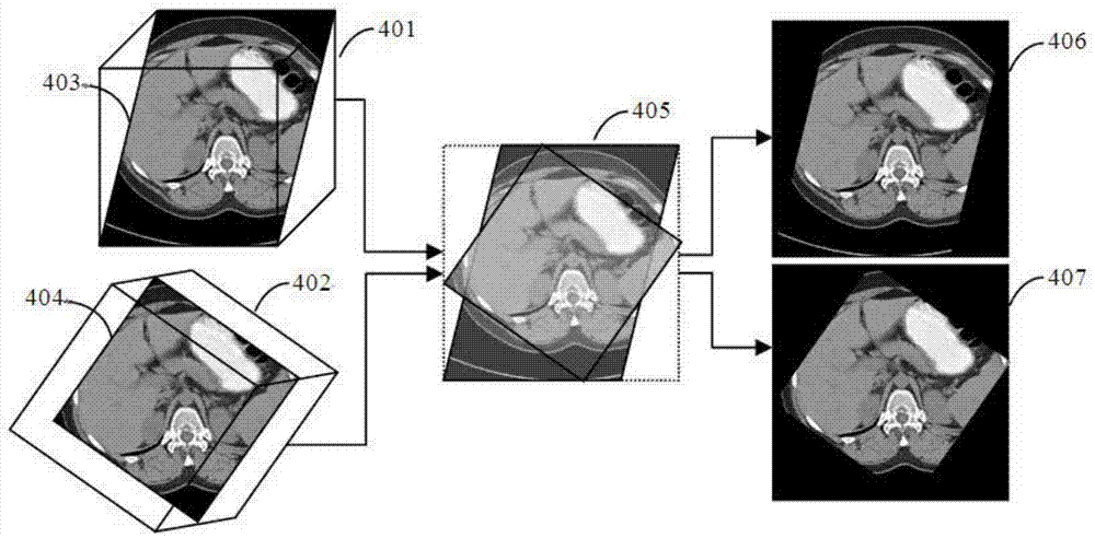
[0107] In this embodiment, the multiple channels of image data are two sets of two-dimensional image data. Using the analysis method of this embodiment, a relatively clear tissue and organ in one channel of data is segmented, and the target area is registered and mapped to In another two-dimensional image, the outline of the target area is displayed in the window by outline, or in the form of a translucent mask. At this time, since there is only a single image, it is not necessary to set multiple sub-windows. It can be displayed in a large window.


PUM


Abstract

The invention provides a medical image analysis method, a medical image analysis system and a medical device. The method comprises the following steps that: a plurality of paths of image data are imported; the spatial coordinate positions of the imported different paths of image data are registered, and registration relations are established; one or more paths of image data are selected, and one or more target regions are separated from each path of image data; the features of the target regions are extracted, and the target regions are mapped into other images according to the registration relations; any spatial plane position is specified, a plurality of section images are generated according to the registered images; and different section position image are correspondingly displayed in different sub-windows, and the same section position images from different image data are displayed in the same sub-windows. With the method of the invention adopted, the technical problems of incapability of embodying the volumetric characteristics of three-dimensional image data and being adverse to the comparison of the differences of different images of a display mode in the prior art can be solved, the subtle differences of images of the same mode or different modes and spatial corresponding position relations of target tissue structures in different modes and time-phase images can be fast and effectively observed, and therefore, image contrast analysis can be facilitated.

Description

technical field [0001] The invention relates to a medical image analysis method, system and medical equipment, belonging to the technical field of medical images. Background technique [0002] At present, for the needs of clinical diagnosis, patients in the medical imaging examination will collect and examine the images of various medical imaging equipment (such as CT, MRI, ultrasound, etc.) Acquisition inspections (such as ultrasound contrast images, tissue maps), and regular inspections of images collected in different periods of the same modality (such as before and after surgery, different stages of the disease cycle), for example, during the treatment of tumors, whether it is surgery Whether it is minimally invasive interventional therapy, regular review of the growth of the lesion is required, and imaging of the target tumor area needs to be collected each time. During this process, the main analysis and evaluation method of tumor growth status is simply measuring the...

Claims


Application Information

Patent Timeline
IPC IPC(8): G06T7/11G06T7/30G06T3/00
CPCG06T3/0068G06T7/0012G06T7/0016G06T2207/20221G06T2207/30096G06T2207/10081
Inventor 孙腾谢晓燕郝永富丛龙飞黄光亮张晓儿
Owner SHENZHEN MINDRAY BIO MEDICAL ELECTRONICS CO LTD
Why Eureka
Try Eureka