Confocal blood vessel imaging machine

A blood vessel imaging and confocal technology, applied in the field of confocal-based blood vessel imaging instrument, can solve the problem of not being able to provide longitudinal position information of blood vessels

Active Publication Date: 2017-03-22
SUZHOU INST OF BIOMEDICAL ENG & TECH CHINESE ACADEMY OF SCI
View PDF16 Cites 5 Cited by
  • Summary
  • Abstract
  • Description
  • Claims
  • Application Information

AI Technical Summary

Problems solved by technology

[0003] Existing CCD-based near-infrared imaging technology can only provide lateral positi

Method used


Image

  • Confocal blood vessel imaging machine

Examples


Embodiment Construction

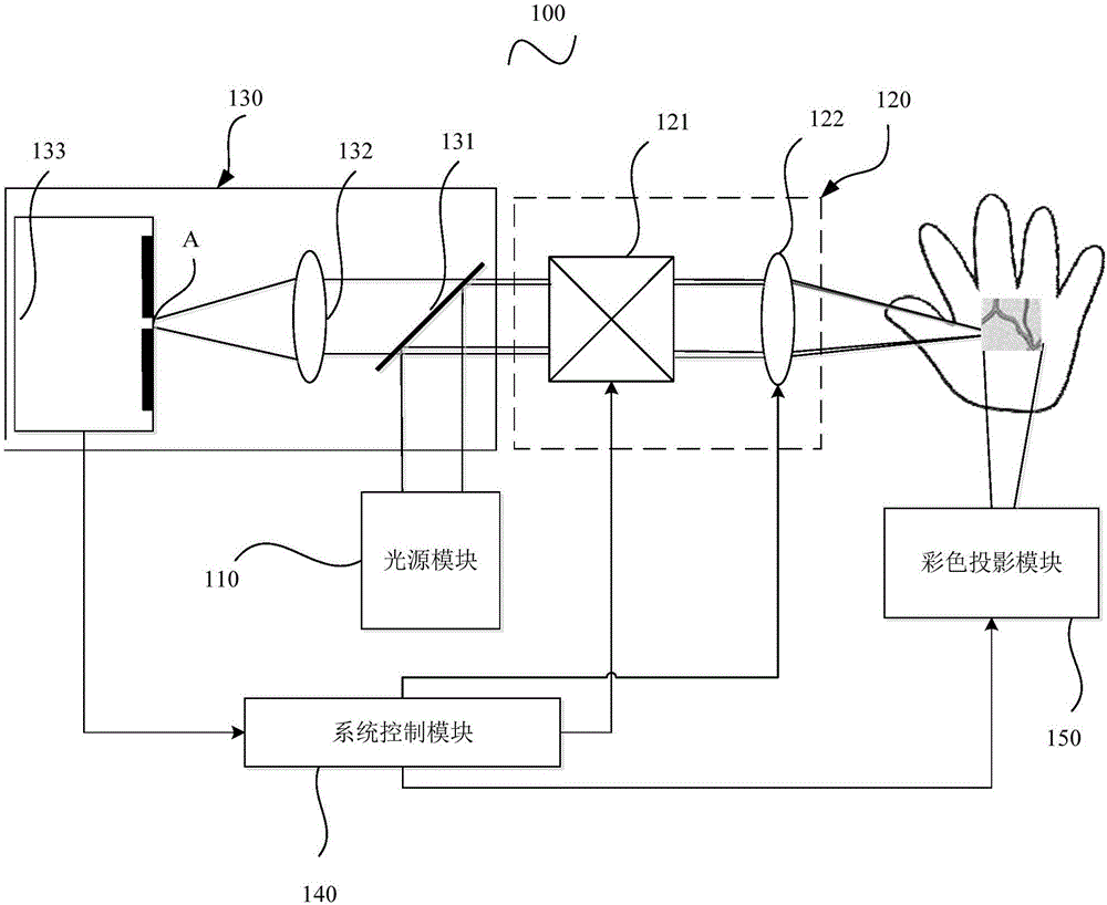
[0020] Please refer to figure 1 A schematic structural diagram of a confocal-based blood vessel imaging apparatus 100 provided for an embodiment of the present invention includes: a light source module 110 , a confocal scanning module 120 , a detection module 130 , a system control module 140 and a color projection module 150 .

[0021] Preferably, the light source module 110 includes a near-infrared light source. What needs to be explained here is that, unlike the near-infrared light source used in the traditional vascular imaging instrument, the illumination method used in the traditional vascular imaging instrument is surface illumination, while in the confocal imaging method provided by the invention, the illumination method used is point illumination .

[0022] It can be understood that the near-infrared light source is used for the illumination light in the process of imaging subcutaneous blood vessels, and in the process of confocal scanning imaging, the difference in ...


PUM


Abstract

The invention provides a confocal blood vessel imaging machine, which comprises a light source module, a confocal scanning module, a detecting module, a color projection module and a system control module. The confocal scanning module comprises an XY light beam scanning mechanism and a scanning lens mechanism; and the detecting module comprises a detector with a pinhole, a focusing lens and a light splitting mechanism. The confocal blood vessel imaging machine provided by the invention utilizes lossless optical sectioning strength of a confocal technology for providing three-dimensional position information of blood vessels, can better provide blood vessel information of obese patients and infant patients for medical personnel, and is beneficial to providing convenience for the medical personnel to give an acupuncture treatment, relieving the pains of the patients, improving the success rate of puncture, shortening the puncture time, reducing the working intensity of the medical personnel, reducing the medical disputes and improving the hospital prestige.

Description

technical field [0001] The invention relates to the field of design and manufacture of a blood vessel imaging instrument, in particular to a confocal-based blood vessel imaging instrument. Background technique [0002] With the improvement of people's living standards, the number of obese patients continues to increase, and infants and ICU patients have high precision requirements for medical staff's needle puncture techniques, making the relationship between doctors and patients inherently fragile. Needle pricks, or even no pricks, become very sensitive. Utilizing the principle that the hemoglobin in blood vessels has a different absorption rate of near-infrared light than other tissues, the in-situ projection of subcutaneous blood vessels is displayed on the skin surface. At present, a "projection infrared vascular imaging instrument" has appeared on the market, using near-infrared image sensors. (Charge-coupled Device, CCD) images blood vessels, and uses visible light to...

Claims


Application Information

Patent Timeline
IPC IPC(8): A61B5/00
CPCA61B5/0068A61B5/0073A61B5/489
Inventor 黄维张运海缪新薛晓君
Owner SUZHOU INST OF BIOMEDICAL ENG & TECH CHINESE ACADEMY OF SCI
Why Eureka
Try Eureka