Use of biological marker detection reagent in preparing kit for detecting invasiveness of pituitary adenomas
A technology of biomarkers and detection reagents, applied in the field of biomedicine, can solve problems such as difficult to achieve total resection
- Summary
- Abstract
- Description
- Claims
- Application Information
AI Technical Summary
Problems solved by technology
Method used
Image


Examples
Embodiment 1
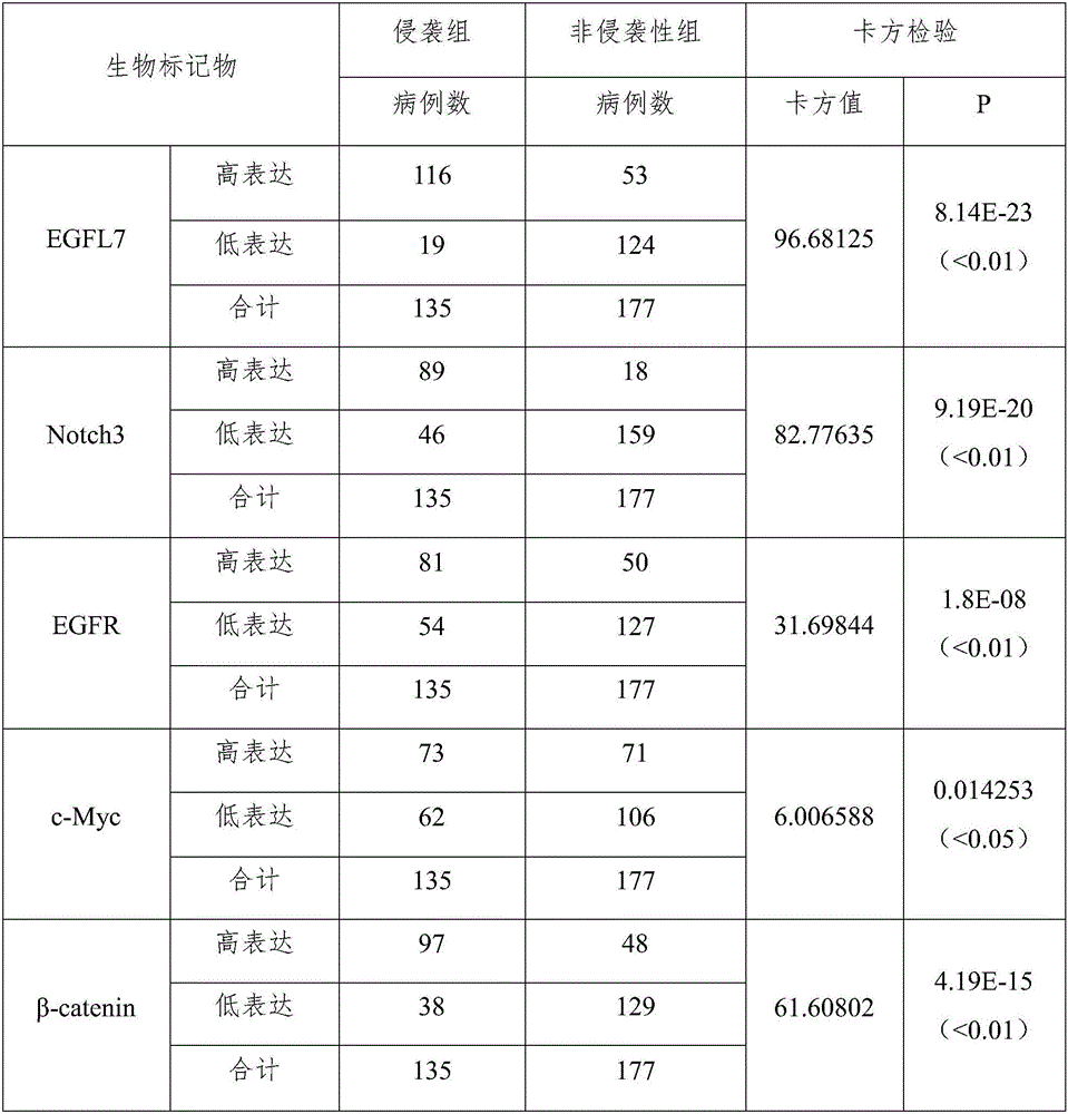
[0039] The clinical data of 312 cases of pituitary adenomas were collected and divided into 135 cases in the invasion group and 177 cases in the non-invasive group according to the Knosp classification. Construct a tissue chip to detect the expression levels of EGFL7, Notch3, EGFR, β-catenin and c-Myc protein in pituitary adenoma specimens using the Leica-BOND III system to perform fully automated immunohistochemical methods, and analyze the above-mentioned proteins and pituitary adenoma invasion, etc. Correlation of clinicopathological characteristics.
[0040] Use Leica automatic scanner to scan, analyze after scanning, give high and low expression interpretation. When the proportion of EGFL7, Notch3, EGFR, c-Myc, β-catenin expressing color 2 points and 3 points respectively reaches 51%-100%, it is determined as high expression, and low expression is the proportion of cells expressing color 1 point 1-100% or 2 points and 3 points are less than 50% of the cells.
[0041] The res...
Embodiment 2
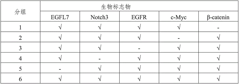
[0046] The method of Example 1 was used to detect the expression level of biomarkers in the 312 pituitary adenoma specimens (including 135 invasive specimens and 177 non-invasive specimens) in Example 1. The only difference is that each specimen was performed 6 times Parallel detection, the types of biomarkers detected each time are shown in Table 2.
[0047] Table 2
[0048]
[0049] In the first group, specimens with high expression of EGFL7, Notch3, EGFR, and c-My were identified as the invasion group, and 74 cases of invasion were detected.
[0050] In the second group, the specimens with high expression of EGFL7, Notch3, EGFR and β-catenin were identified as invasive specimens, and a total of 99 invasive specimens were detected.
[0051] In the third group, the specimens with high expression of EGFL7, Notch3, c-My and β-catenin were identified as invasive specimens, and 77 invasive specimens were detected.
[0052] In the fourth group, the specimens with high expression of EGFL7, ...
Embodiment 3
[0057] In this example, the Transwell experiment was used to identify the invasion ability of pituitary adenoma cells.
[0058] 1. Melt BD Matrigel as a liquid reserve.
[0059] 2. Dilute Matrigel with serum-free medium at a volume ratio of 1:8 (Matrigel: serum-free medium).
[0060] 3. Add 80μL of the diluted Matrigel working solution to the upper chamber of the Transwell chamber, and incubate it in an incubator at 37°C for 6 hours to polymerize the Matrigel gel.
[0061] 4. When the pituitary adenoma cells (GH3, purchased from ATCC) grow to a confluence rate of 85%, when they are in good condition, digest the cells, resuspend the cells in serum-free medium, and count and adjust the density to 2×10 5 Cells / ml to obtain cell suspension.
[0062] 5. Including five treatment groups, among which treatment groups 1-3: add about 200μl of the above cell suspension in the upper chamber of the Transwell chamber (24-well plate, 8-plate), and divide it into 3 different treatment groups to add the...
PUM
 Abstract
Description
Claims
Application Information
 - R&D
- Intellectual Property
- Life Sciences
- Materials
- Tech Scout
- Unparalleled Data Quality
- Higher Quality Content
- 60% Fewer Hallucinations
Browse by: Latest US Patents, China's latest patents, Technical Efficacy Thesaurus, Application Domain, Technology Topic, Popular Technical Reports.
© 2025 PatSnap. All rights reserved.Legal|Privacy policy|Modern Slavery Act Transparency Statement|Sitemap|About US| Contact US: help@patsnap.com