Noninvasive circulating tumor cell detection and diagnosis device and detection and diagnosis method thereof
A technology for diagnosing devices and tumor cells, which is applied in the fields of basic medical scientific research and clinical medical testing instruments and equipment, can solve the problems of inaccurate detection results and affect the physiological environment of cells, etc., and achieves high display quality, simple device structure, and low power consumption. Effect
Inactive Publication Date: 2016-02-03
周辉
View PDF2 Cites 3 Cited by
- Summary
- Abstract
- Description
- Claims
- Application Information
AI Technical Summary
Problems solved by technology
In vivo fluorescent labeling flow cytometry developed in recent years overcomes the problem that imaging detection cannot detect circulating tumor cells in the blood of cancer patients early, and provides a new detection method for early detection of c
Method used
View moreImage
Smart ImageViewing Examples
Examples
 PUM
| Property | Measurement | Unit |
|---|---|---|
| Wavelength | aaaaa | aaaaa |
Abstract
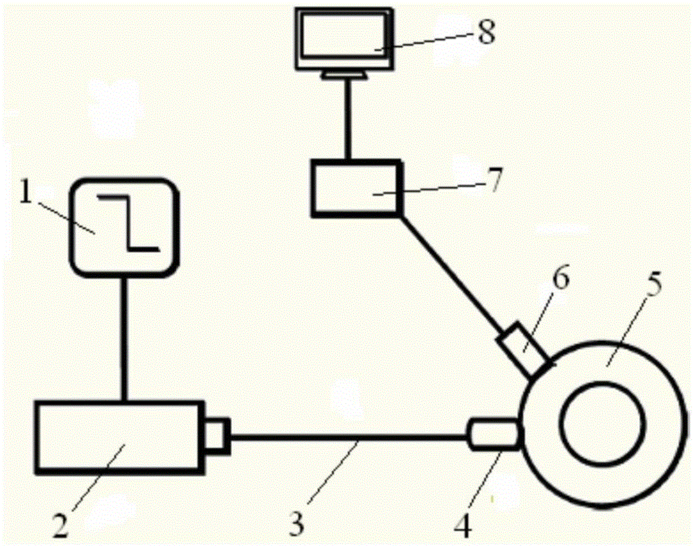
The invention discloses a noninvasive circulating tumor cell detection and diagnosis device. A high-frequency impulse laser (2), a light beam coupler (4) and an ultrasonic probe (6) are arranged, and the light beam coupler (4) and the ultrasonic probe (6) are both attached to a skin contactor (5). The invention further discloses a detection and diagnosis method of the detection and diagnosis device. According to the technical scheme, on the basis of an in-vivo fluorescence detection method, a photoacoustic effect of biological tissue is combined, and the inaccuracy caused by environmental changes brought by ex-vivo detection is overcome by utilizing photoacoustic specificity of the biological tissue and the deep penetrability of an ultrasonic signal; the effects of clinical detection and noninvasive detection are achieved, blood drawing and marking are not needed, and a novel approach is provided for early detection of clinical tumor cells; the advantages of in-vivo and real-time monitoring are achieved, and detection can be conducted on a patient for a long time; the device is wide in purpose, the equipment structure is simple, operation is convenient, and the safety is high.
Description
technical field [0001] The invention belongs to the technical field of basic medical scientific research and clinical medical testing equipment. More specifically, the present invention relates to a non-invasive detection and diagnosis device for circulating tumor cells. In addition, the present invention also relates to its detection and diagnosis method. Background technique [0002] Cancer is a disease that seriously threatens human health and life. If cancer patients can be detected and treated in time, not only the survival rate can be improved, but also the quality of life of patients can be improved. Therefore, early detection, early diagnosis and timely treatment of cancer are necessary. [0003] In recent years, the early diagnosis of cancer includes imaging diagnosis such as: photoacoustic imaging, photoacoustic tomography, photoacoustic spectral microscopy imaging, etc. The results that can be detected by imaging are often in the middle and late stages of cancer ...
Claims
Login to View More Application Information
Patent Timeline
 IPC IPC(8): A61B5/00A61B8/00
Inventor 周辉
Owner 周辉
Features
- Generate Ideas
- Intellectual Property
- Life Sciences
- Materials
- Tech Scout
- Unparalleled Data Quality
- Higher Quality Content
- 60% Fewer Hallucinations
Learn More Browse by: Latest US Patents, China's latest patents, Technical Efficacy Thesaurus, Application Domain, Technology Topic, Popular Technical Reports.
© 2025 PatSnap. All rights reserved.Legal|Privacy policy|Modern Slavery Act Transparency Statement|Sitemap|About US| Contact US: help@patsnap.com

