
System and apparatus of digital medical image for X-ray system

A technology for medical imaging and X-rays, which is applied to instruments for radiological diagnosis, radiation detection devices, medical images, etc., and can solve problems such as difficulty in setting up bridge cables, monitoring of emission time, and difficulty in laying cables.

Inactive Publication Date: 2015-10-07
申东埈
View PDF6 Cites 2 Cited by
  • Summary
  • Abstract
  • Description
  • Claims
  • Application Information

AI Technical Summary

Problems solved by technology

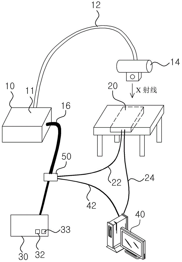
Therefore, it is difficult to lay the corresponding cables 22, 42 for transmitting the control signals output by the controller 30 to the generator 10 to the X-ray detector 20 and / or the monitoring device 40
Moreover, usually the company that produces or supplies the X-ray system and the digital medical imaging system is not the same company, so it is difficult to set up a separate bridge device and corresponding cables 22, 42
In addition, the types of control signals used by the X-ray systems of each manufacturer are different, so it is difficult to install a DR (Digital Medical Imaging) system and software for the system that match the corresponding X-ray system
[0010] In addition, according to the existing method, the monitoring device 40 only receives control signals such as the preparation signal and the emission signal of the controller 30, so only the control signals and the X-ray image information received from the X-ray detector 20 are monitored, and Unable to monitor other X-ray emission information (for example, X-ray intensity, emission time, emission amount, etc.)

Method used


Image

  • System and apparatus of digital medical image for X-ray system
  • System and apparatus of digital medical image for X-ray system
  • System and apparatus of digital medical image for X-ray system

Examples


Embodiment Construction

[0036] The present invention can be modified in many ways, and can have various embodiments. Specific embodiments are shown in the drawings and described in detail in the description below. However, the present invention is not limited to specific embodiments, and it should be understood that the present invention also includes all changes, equivalents and substitutions that do not depart from the technical idea and technical scope of the present invention.

[0037] Hereinafter, embodiments according to the present invention will be described in detail with reference to the drawings. When describing the drawings, the same or corresponding components will be assigned the same reference numerals regardless of the reference numerals, and redundant descriptions will be omitted.

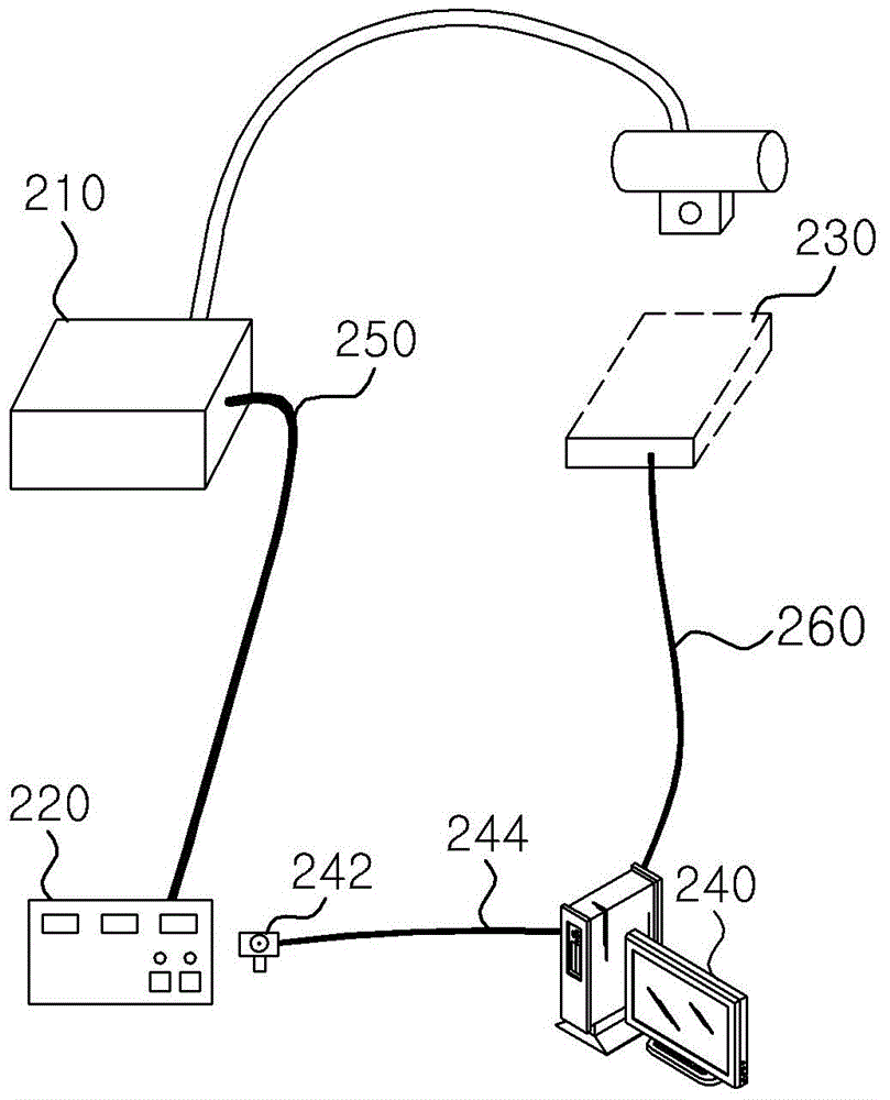
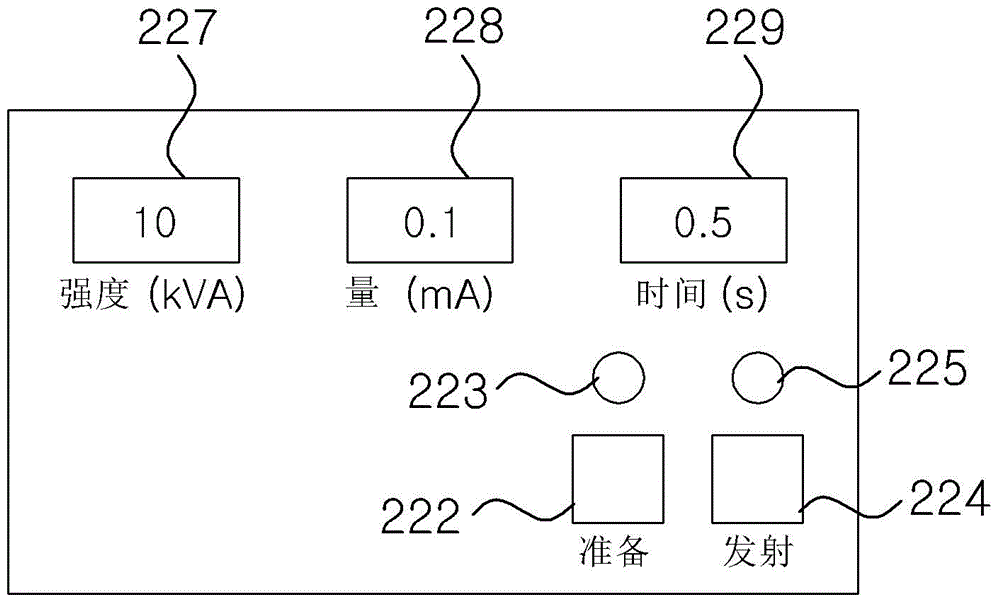
[0038] figure 2 In order to show the composition diagram of the digital medical imaging system used for X-ray system related to an embodiment of the present invention, image 3 To show the front view of...


PUM


Abstract

Digital radiography system and device for an X-ray system are provided. The digital radiography system includes: an X-ray detector that receives X-rays radiated from a generator of an X-ray system and that forms an X-ray image; a camera that captures an image of one side surface of a controller outputting a ready signal and a shot signal to control operation of the generator; and a monitoring device that analyzes image information transmitted from the camera to recognize a control signal output from the controller to the generator and that provides information on the control signal to the X-ray detector. By acquiring a control signal of the generator using an external image of the controller using the camera, it is not necessary to connect a cable for allowing the monitoring device and the X-ray detector to directly receive the control signal from the controller and thus to improve convenience of installation.

Description

technical field [0001] The present invention relates to a digital medical imaging (DR: digital radiography) system and device, in particular to a digital medical imaging system and device for an X-ray system, which uses a camera to identify and process the actions of the controller of the X-ray generator of the X-ray system . Background technique [0002] Nowadays, with the development of science and medical technology, the field of radiation-related equipment in diagnostic equipment has achieved rapid development. For most patients, radiation examination has become a very common diagnostic method. The most popular diagnostic equipment in radiation-related equipment is X-ray system, which is used to view anatomical structures and detect pathological phenomena, diseases or abnormal anatomical structures. [0003] figure 1 In order to summarize and display the composition diagram of the existing digital medical imaging system for X-ray system. [0004] refer to figure 1 , ...

Claims


Application Information

Patent Timeline
Patent Type & Authority Applications(China)
IPC IPC(8): A61B6/00
CPCA61B6/54A61B6/40A61B6/42A61B6/462A61B6/548G06F19/3406G16H30/20G16H40/63A61B6/563
Inventor 申东埈
Owner 申东埈
Learn More