
Myasthenia gravis detection kit with non-coding RNA Tmevpg1 as detection or diagnosis screening marker and application of kit

A myasthenia gravis and detection kit technology, applied in the biological field, can solve problems such as no research reports

Active Publication Date: 2015-04-29
XIANGYA HOSPITAL CENT SOUTH UNIV
View PDF0 Cites 3 Cited by
  • Summary
  • Abstract
  • Description
  • Claims
  • Application Information

AI Technical Summary

Problems solved by technology

However, there is no research report on the role of Tmevpg1 in myasthenia gravis.

Method used


Image

  • Myasthenia gravis detection kit with non-coding RNA Tmevpg1 as detection or diagnosis screening marker and application of kit
  • Myasthenia gravis detection kit with non-coding RNA Tmevpg1 as detection or diagnosis screening marker and application of kit
  • Myasthenia gravis detection kit with non-coding RNA Tmevpg1 as detection or diagnosis screening marker and application of kit

Examples


Embodiment 1

[0021] 1. RNA extraction method:

[0022] (1) Take 5ml of peripheral cubital vein blood sample with EDTA anticoagulant blood collection tube.

[0023] (2) Centrifuge at 3000 rpm for 10 min at room temperature, and carefully suck out the upper serum.

[0024] (3) Add an equal volume of PBS (1×) solution to the remaining blood sample, and fully blow and mix to dilute the blood sample;

[0025] (4) Slowly add the above-mentioned diluted blood sample to another centrifuge tube that has been added with an equal volume of lymphocyte separation liquid against the wall, and keep the above-mentioned mixed liquid above the liquid level of the lymphocyte separation liquid (that is, the two liquids Do not mix, keep a clear interface), centrifuge at 2400rpm for 20min at room temperature;

[0026] (5) Use a pipette gun to carefully absorb the second white blood cell membrane layer in the centrifuged specimen, put it into a new enzyme-free centrifuge tube, centrifuge at 3000rpm, 4°C for 10...


PUM


Abstract

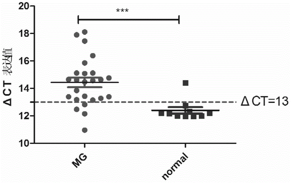
The invention discloses a myasthenia gravis detection kit with non-coding RNA Tmevpg1 as a detection or diagnosis screening marker and an application of the kit. The kit is applicable to myasthenia gravis screening and diagnosis by relatively quantitatively detecting the expression level of Tmevpg1 in the peripheral blood specimen of a myasthenia gravis patient.

Description

technical field [0001] The invention belongs to the field of biotechnology, and specifically relates to a myasthenia gravis detection kit and application using non-coding RNA Tmevpg1 as a detection or diagnostic screening marker. Background technique [0002] Myasthenia gravis (MG) is a B-cell-mediated, T-cell-dependent, and complement-involved autoimmune disease involving the neuromuscular junction, and its clinical manifestations are neuromuscular junction (NMJ) transmission disorders Resulting skeletal muscle weakness and fatigue. Studies have shown that the anti-acetylcholine receptor (acetylcholine receptor, AchR) antibody at the NMJ is the main antibody, and about 40% of anti-AchR negative systemic MG patients can be anti-MuSK (muscle-specific tyrosine kinase,) antibody positive, anti - About 20% of systemic MG patients with negative AchR and anti-MuSK antibodies may be positive for anti-LRP4 (Lipoprotein-Related Protein 4) antibodies. The pathogenesis of MG is relat...

Claims


Application Information

Patent Timeline
IPC IPC(8): C12Q1/68
CPCC12Q1/6883C12Q2600/158
Inventor 罗朝辉刘晓芳罗梦川杨欢肖波徐立群李秋香
Owner XIANGYA HOSPITAL CENT SOUTH UNIV
Learn More