Control method of X-ray photographing system

A control method and X-ray technology, which is applied in the fields of radiological diagnostic instruments, medical science, and diagnosis, and can solve problems such as rack errors, ray fields exceeding the receiving range of flat-panel detectors, and parameter changes.

Active Publication Date: 2015-03-25
SHANGHAI UNITED IMAGING HEALTHCARE
View PDF10 Cites 8 Cited by
  • Summary
  • Abstract
  • Description
  • Claims
  • Application Information

AI Technical Summary

Problems solved by technology

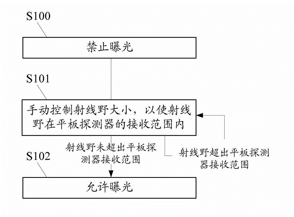
[0007] A control method of an X-ray imaging system in the prior art, such as figure 2 As shown, the following steps are included to solve the problem that the ray field in X-ray photography exceeds the receiving range of the flat panel detector:
[0018] The manual adjustment of the ray field in the prior art is controlled by experience or intuition, which may cause changes in the parameters of the source image distance and other racks during operation, resulting in other errors and accuracy problems in the rack operation

Method used


Image

  • Control method of X-ray photographing system
  • Control method of X-ray photographing system
  • Control method of X-ray photographing system

Examples


Embodiment Construction

[0111] In order to automatically prevent the ray field from exceeding the receiving range of the flat panel detector plane during X-ray photography, so as to better guide the operation of the X-ray photography system, this embodiment provides a control method for the X-ray photography system, such as image 3 shown, including:

[0112] Step S200, acquiring rack information.

[0113] In this step, the frame of the X-ray photography system includes a tube for emitting X-rays and a flat panel detector for receiving the X-rays. For its specific structure, reference may be made to the prior art. This embodiment does not modify the X-ray photography system. The frame is limited.

[0114] The concept of the frame information can be broadly understood: in the process of X-ray photography, all kinds of operating information output or generated by the circuits and sensing nodes of the frame can be considered as the frame information; the frame manual, instruction manual or The inheren...


PUM


Abstract

The invention relates to a control method of an X-ray photographing system. The control method includes the steps of obtaining rack information and obtaining the scope of a plane of a flat panel detector; according to the rack information, calculating a radiation field generated by X-rays on the plane of the flat panel detector through a beam-defining clipper; when the radiation field exceeds the scope of the plane of the flat panel detector, adjusting at least one parameter of the width or the height of the radiation field so as to enable the adjusted radiation field to be smaller than or equal to the scope of the plane of the flat panel detector; and transmitting the adjusted parameter to the beam-defining clipper. By means of the control method, the X-ray photographing system can automatically solve the problem that the radiation field exceeds the scope of the plane of the flat panel detector in the X-ray photographing.

Description

technical field [0001] The invention relates to an X-ray photography system, in particular to a control method of the X-ray photography system. Background technique [0002] The essence of X-ray is a ray with a short wavelength but can penetrate the human body. It is used in medicine as a medical X-ray photography system, referred to as X-ray photography system, and it is also one of the six major medical imaging equipment. X-ray photography system is the routine equipment and necessary equipment in the radiology department and orthopedics department of the hospital. Since the development of medical X-ray imaging technology, the variety and output of X-ray imaging systems have greatly increased. At present, high-frequency, digital, and multi-functional X-ray imaging systems have become the mainstream of the market. [0003] see figure 1 The frame structure of an X-ray photography system shown mainly includes a mounting beam 1, a photography bed 2, a flat detector (FD) 3, a...

Claims


Application Information

Patent Timeline
IPC IPC(8): A61B6/00
Inventor 丁健
Owner SHANGHAI UNITED IMAGING HEALTHCARE
Why Eureka
Try Eureka