An automatic detection method of vascular adventitia based on intravascular ultrasound images

An ultrasound image and vascular adventitia technology, applied in the field of medical image processing, can solve the problems of reducing the accuracy of statistical modeling, complex method models, etc., to achieve the effect of ensuring automation and avoiding complexity

Active Publication Date: 2018-02-27
SUZHOU INST OF BIOMEDICAL ENG & TECH CHINESE ACADEMY OF SCI
View PDF3 Cites 0 Cited by
  • Summary
  • Abstract
  • Description
  • Claims
  • Application Information

AI Technical Summary

Problems solved by technology

At present, there are three main types of computer automatic detection (segmentation) algorithms for blood vessel edges based on IVUS images: the first type is statistical methods (G. Mendizabal-Ruiz, M. Rivera, et al., “Aprobabilistic segmentation method for the identification of luminal borders in intravascular ultrasound images", IEEE Conference on Computer Vision and Pattern Recognition, pp.1-8, 2008.), statistical modeling of the gray distribution of the image to achieve IVUS image segmentation, so as to detect the edge of the blood vessel, but in the IVUS image Complex image features such as artifacts and calcifications will greatly reduce the accuracy of statistical modeling; the second category is machine learning methods (1.E. G. Bovenkamp, ​​J. Dijkstra, J. G.Bosch, et al., “Multi-agent segmentation of IVUS images”, Patten Recognition, Vol.37, No.4, pp.647-663, 2004; 2. G. Unal, S. Bucher, S. Carlier, et al., “Shape-driven segmentation of the arterial wall in intravascular ultrasoundimages", IEEE Trans. On information technology in biomedicine, Vol.12, No.3, pp.335-346, 2008.), this type of method model is complicated, and it is subject to many restrictions in practical application; the third type is Method based on active contour model (1. Zhang Qi, Wang Yuanyuan, etc., "Active Contour Model and Contourlet Multi-resolution Analysis to Segment Intravascular Ultrasound Images", Optical Precision Engineering, Vol.16, No.11, pp.2301- 311, 2008; 2. X. Zhu, P.Zhang, J. Shao, et al., “A snake-based method for segmentation of intravascular ultrasound images and its in vivo validation”, Ultrasonics, Vol.51, pp.181-189, 2011.), this type of method is simple and easy to implement, but it often needs to give an initial contour line, and the detection results are easily affected by complex image features such as noise

Method used


Image

  • An automatic detection method of vascular adventitia based on intravascular ultrasound images
  • An automatic detection method of vascular adventitia based on intravascular ultrasound images
  • An automatic detection method of vascular adventitia based on intravascular ultrasound images

Examples


Embodiment Construction

[0021] The present invention will be further described below in conjunction with examples of implementation and accompanying drawings, but the protection scope of the present invention should not be limited by this.

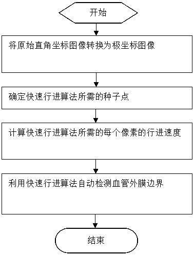
[0022] figure 1 It is a flow chart of an automatic detection method for the adventitia of a blood vessel based on an intravascular ultrasonic image of the present invention. As shown in the figure, an automatic detection method of vascular adventitia based on intravascular ultrasound (IVUS: Intravascular Ultrasound) images includes a process of converting intravascular ultrasound images from rectangular coordinates to polar coordinates; The process of seed points required by the Marching algorithm; including a process of determining the speed of travel at each pixel required by the Fast Marching algorithm according to the image grayscale and gradient; including an automatic detection using the Fast Marching algorithm The process of the adventitia of blood vessel...


PUM


Abstract

The invention discloses an intravascular ultrasound image based automatic adventitia detection method. The method includes processes: transforming an intravascular ultrasound image from rectangular coordinates to polar coordinates; determining seed points needed by a Fast Marching algorithm; determining marching speed of each pixel needed by the Fast Marching algorithm according to image grayscale and gradient; automatically detecting the adventitia by the aid of the Fast Marching algorithm. The seed points, a termination point and a valid marching speed function are detected automatically, so that automaticity in the detection process is guaranteed; by the Fast Marching algorithm based processing method, simpleness and effectiveness of the detection method are guaranteed, and complexity of an existing algorithm model and dependence on imaging conditions are avoided.

Description

technical field [0001] The present invention relates to the field of medical image processing, in particular to a Fast Marching algorithm and an automatic detection method for intima and intima of blood vessels applied to intravascular ultrasound (IVUS: Intravascular ultrasound) images. Background technique [0002] Intravascular Ultrasound (IVUS: Intravascular Ultrasound) images have very important clinical application value for the diagnosis and treatment of cardiovascular diseases such as atherosclerosis. Diagnosis of atherosclerosis based on IVUS images requires quantitative indicators of atherosclerotic image features such as vessel lumen area and plaque area. The accurate extraction of these quantitative indicators depends on effective vessel edge detection. Manual detection means that doctors manually outline the lumen and adventitia boundaries of blood vessels, which is not only time-consuming and laborious, but also limited by the subjectivity of doctors' experience...

Claims


Application Information

Patent Timeline
Patent Type & Authority Patents(China)
IPC IPC(8): G06T7/13
CPCG06T3/604G06T7/0012G06T2207/10132G06T2207/30101
Inventor 严加勇向永嘉崔崤峣简小华韩志乐
Owner SUZHOU INST OF BIOMEDICAL ENG & TECH CHINESE ACADEMY OF SCI
Why Eureka
Try Eureka