
A method for quantitatively detecting the amount of carbon dioxide exhaled by cancer cells

A technology for quantitative detection of carbon dioxide, applied in the field of fluorescent biosensors, can solve the problems of not being able to monitor the metabolic process of cancer cells and not having the ability to quantitatively detect carbon dioxide in cells

Inactive Publication Date: 2016-11-23
BEIJING INSTITUTE OF TECHNOLOGYGY
View PDF5 Cites 0 Cited by
  • Summary
  • Abstract
  • Description
  • Claims
  • Application Information

AI Technical Summary

Problems solved by technology

[0005] Aiming at the problem that the fluorescent probes currently used for detecting the metabolic process of cancer cells are used for live cell imaging, there is no quantitative detection of carbon dioxide produced by cells, and the problem that the metabolic process of cancer cells cannot be monitored. The purpose of the present invention is to provide a method for quantitative detection of cancer cells. Method for exhaled carbon dioxide content by cells using a method containing (2-dimethylamino)ethyl 4,4',4"-(1H-pyrrole-1,2,5-triyl)tribenzoate (TPP -DMAE) fluorescent probe detects the metabolic process of living cells. The fluorescent probe is used for fluorescence imaging of cancer cells and can quantitatively detect the carbon dioxide content produced by the metabolism of cancer cells, and realize the identification of cancer cell types

Method used


Image

  • A method for quantitatively detecting the amount of carbon dioxide exhaled by cancer cells
  • A method for quantitatively detecting the amount of carbon dioxide exhaled by cancer cells
  • A method for quantitatively detecting the amount of carbon dioxide exhaled by cancer cells

Examples


Embodiment 1

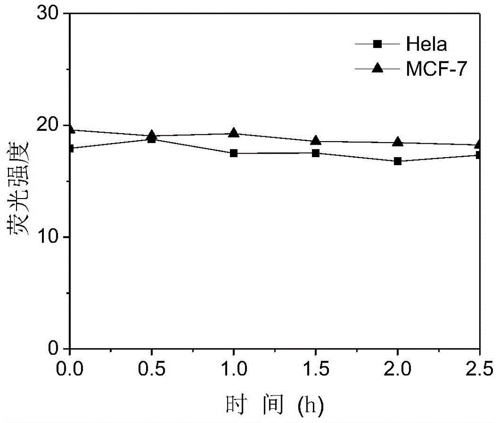
[0043] (1) Dissolve 0.64mg TPP-DMAE in 0.01mL dimethyl sulfoxide (DMSO) to obtain a concentration of 1×10 -2 mol / L solution a; add 10mL high-sugar DMEM medium to solution a to obtain a concentration of 1×10 -5 mol / L solution b.

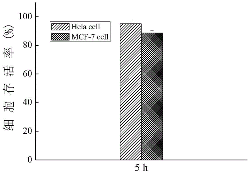
[0044] (2) ① In order to detect whether TPP-DMAE can cause the inactivation of Hela cell and MCF-7cell within the test time range, the following toxicity test experiments were done:

[0045] Cervical cancer cells (Hela cells) and breast cancer cells (MCF-7 cells) were respectively passaged into two culture dishes dedicated to confocal laser microscopy (confocal); the Hela cells, MCF-7cell was digested with 0.05% trypsin until 50-60% of the cells were detached from the bottom of the culture dish, and 3 mL of high-sugar DMEM medium was added to the culture dish of Hela cell and MCF-7 cell to stop the digestion, and then Hela cell Cell and MCF-7cell suspensions were transferred from the petri dish to two 15mL centrifuge tubes, each supplemented with 7m...

Embodiment 2

[0055] (1) Dissolve 0.64mgTPP-DMAE in 0.01mL dimethyl sulfoxide (DMSO) to obtain a concentration of 1×10 -2 mol / L solution a; add 10mL high-sugar DMEM medium to solution a to obtain a concentration of 1×10 -5 mol / L solution b.

[0056] (2) ① In order to detect whether TPP-DMAE can cause the inactivation of Hela cell and MCF-7cell within the test time range, the following toxicity test experiments were done:

[0057] Cervical cancer cells (Hela cells) and breast cancer cells (MCF-7 cells) were respectively passaged into two culture dishes dedicated to confocal laser microscopy (confocal); the Hela cells, MCF-7cell was digested with 0.05% trypsin until 50-60% of the cells were detached from the bottom of the culture dish, and 3 mL of high-sugar DMEM medium was added to the culture dish of Hela cell and MCF-7 cell to stop the digestion, and then Hela cell Cell and MCF-7cell suspensions were transferred from the petri dish to two 15mL centrifuge tubes, each supplemented with 7mL...

Embodiment 3

[0067] (1) Dissolve 0.64mg TPP-DMAE in 0.01mL dimethyl sulfoxide (DMSO) to obtain a concentration of 1×10 -2 mol / L solution a; add 10mL high-sugar DMEM medium to solution a to obtain a concentration of 1×10 -5 mol / L solution b.

[0068] (2) ① In order to detect whether TPP-DMAE can cause the inactivation of Hela cell and MCF-7cell within the test time range, the following toxicity test experiments were done:

[0069] Cervical cancer cells (Hela cells) and breast cancer cells (MCF-7 cells) were respectively passaged into two culture dishes dedicated to confocal laser microscopy (confocal); the Hela cells, MCF-7cell was digested with 0.05% trypsin until 50-60% of the cells were detached from the bottom of the culture dish, and 3 mL of high-sugar DMEM medium was added to the culture dish of Hela cell and MCF-7 cell to stop the digestion, and then Hela cell Cell and MCF-7cell suspensions were transferred from the petri dish to two 15mL centrifuge tubes, each supplemented with 7m...


PUM


Abstract

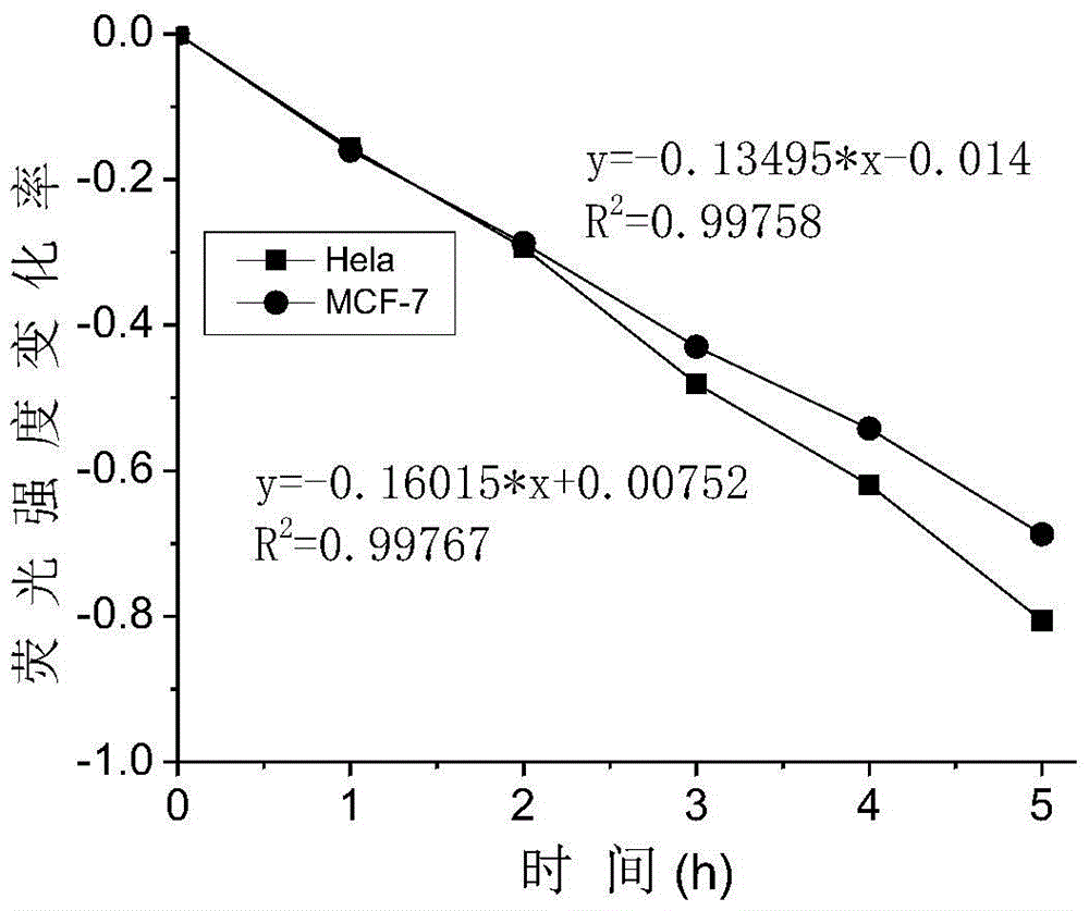
The invention discloses a method applied to quantitative detection of carbon dioxide exhaled by cancer cells, belonging to the field of fluorescent biosensors. The method comprises the following steps: firstly dissolving TPP-DMAE in DMSO and adding a high-glucose DMEM culture medium to obtain a mixed solution; transferring the cancer cells into a culture dish, culturing and washing, adding the mixed solution and the high-glucose DMEM culture medium, culturing and washing, adding a fixation agent, standing, washing, adding a buffer solution to obtain a to-be-detected sample; detecting the fluorescence intensity of the to-be-detected sample, comparing a value of a fluorescence intensity-time change rate caused by CO2 generated by self metabolism of the cancer cells with a value of a fluorescence intensity-gas volume change rate after introducing gas into deactivation cells, and taking the volume of gas introduced in the time period corresponding to the same fluorescence intensity change rate as the volume of CO2 generated by the self metabolism of the cancer cells in different time periods. The method is capable of quantitatively detecting the content of carbon dioxide generated by metabolism of active cells and discriminating the types of the cancer cells.

Description

technical field [0001] The invention relates to a method for quantitatively detecting the content of carbon dioxide exhaled by cancer cells, belonging to the field of fluorescent biosensors. Background technique [0002] Metabolism is the basic condition for organisms to maintain life activities. The characteristics of growth, development, inheritance and variation in the life process are all based on metabolism. The normal activities of the body's physiological and biochemical functions are completely dependent on the normal physiology and metabolism of the cells. When the cells become diseased, the human body will suffer from diseases, and once the metabolism stops, life will end. Therefore, it is of great significance to realize the recording and monitoring of cell metabolism. Among them, active cancer cells are characterized by unlimited and endless proliferation, which consumes a lot of nutrients in the patient's body; cancer cells themselves release a variety of toxin...

Claims


Application Information

Patent Timeline
Patent Type & Authority Patents(China)
IPC IPC(8): G01N21/64
Inventor 董宇平王焕陈笛笛张亚会石建兵佟斌
Owner BEIJING INSTITUTE OF TECHNOLOGYGY
Learn More