A kind of preparation method of insect sample for scanning electron microscope
A scanning electron microscope and insect technology, applied in the field of insect sample preparation, can solve the problems of slow penetration of glutaraldehyde, high degree of image shrinkage, sample deformation, etc., to achieve great practical application value, good insect morphological structure, prevent shrunken effect
- Summary
- Abstract
- Description
- Claims
- Application Information
AI Technical Summary
Problems solved by technology
Method used
Image
Examples
Embodiment 1
[0038] Embodiment 1 (different from the comparative example in step 2)
[0039] The preparation method steps of scanning electron microscope insect sample of the present invention are as follows:
[0040] 1) Gently brush the surface of the larvae of the peach moth borer with an ear washing ball, and then wash the larvae three times with 0.1M phosphate buffer solution (pH7.2);
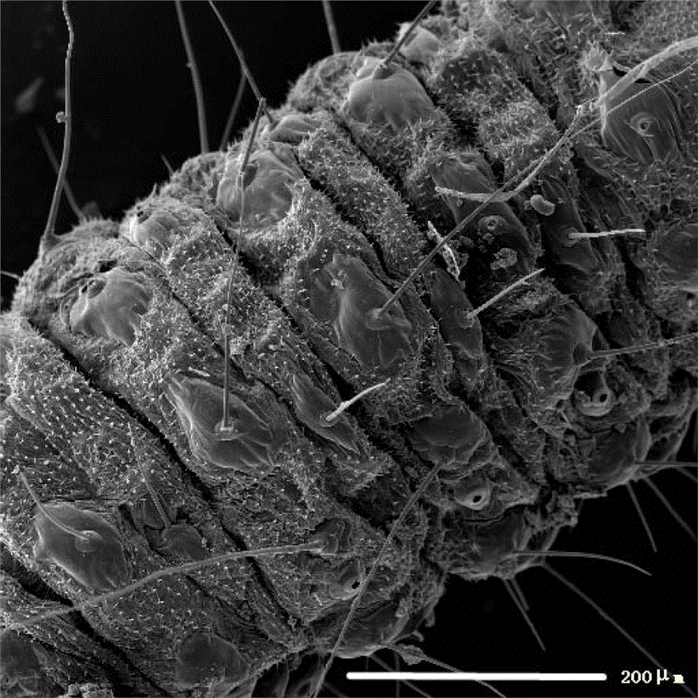
[0041] 2) Immerse the larvae in the fixative solution (fixative solution is 3% glutaraldehyde), lightly prick three small holes on each side of the larvae under a dissecting microscope, and transfer to a 1.5ml centrifuge tube, which contains fixative Liquid about 1ml. Place the centrifuge tube in a centrifuge tube rack and heat it in a microwave oven on high heat for 8 seconds. At this time, the centrifuge tube is hot but not hot. Replace the fixative in the centrifuge tube and repeat the heating twice, then fix in a refrigerator at 5°C for 12 hours;
[0042] 3) Wash the larvae 5 times with phosphate...
Embodiment 2
[0053] The difference between this embodiment and embodiment 1 is:
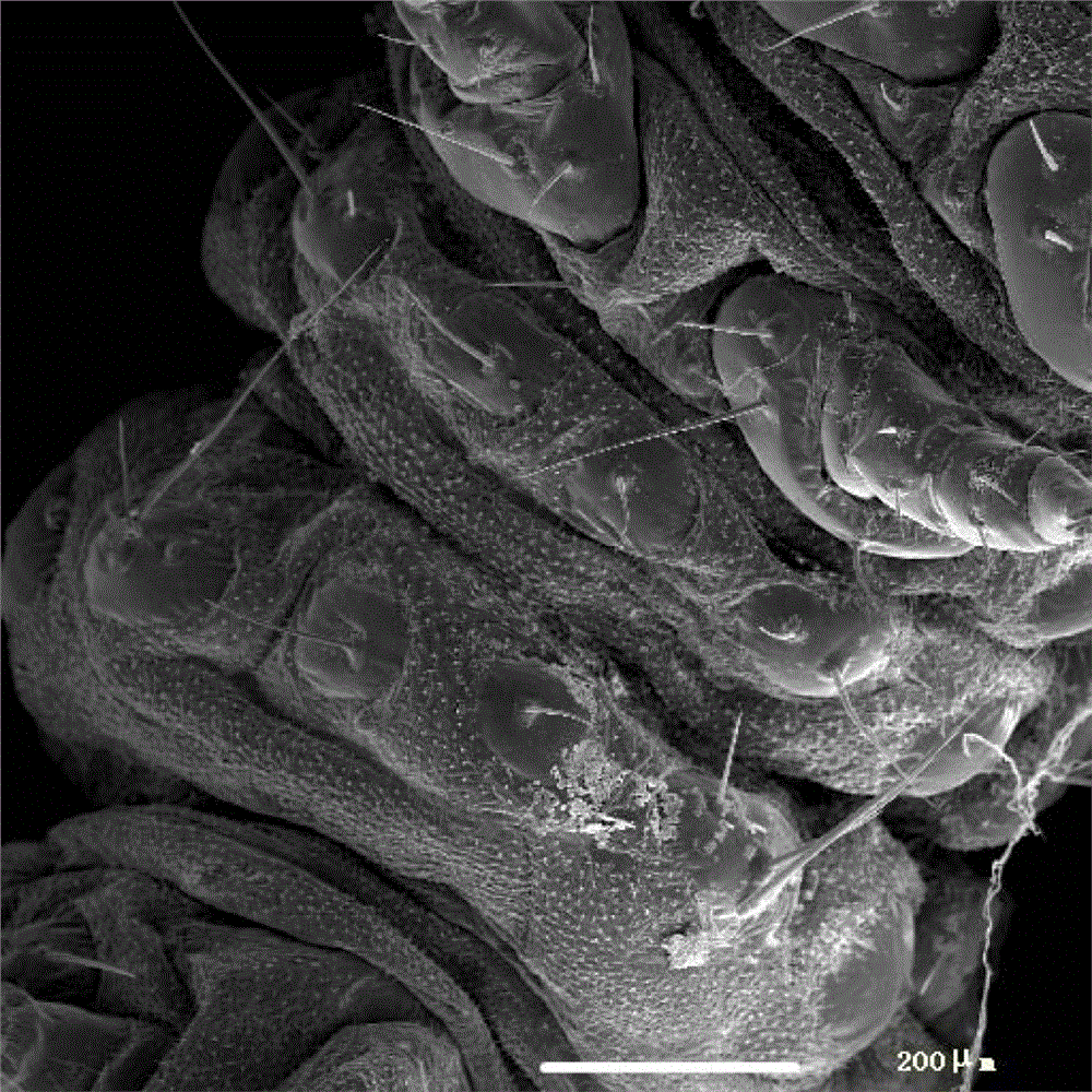
[0054] (2) Immerse the larvae in the fixative solution, which is prepared by adding 12 μl Tween-80 to every 20ml of 2.5% glutaraldehyde, lightly puncture three small holes on each side of the larvae under a dissecting microscope, Transfer to a 1.5ml centrifuge tube with about 1ml of fixative in the centrifuge tube. Place the centrifuge tube in a centrifuge tube rack and heat it in a microwave oven on high heat for 8 seconds. At this time, the centrifuge tube is hot but not hot. Replace the fixative in the centrifuge tube and repeat the heating twice, then fix in the refrigerator at 5°C for 12h.
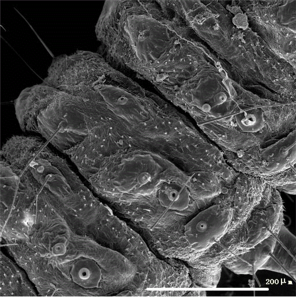
[0055] All the other steps are the same as in Example 1. It can be seen from the imaging results that the abdominal nodes and internodes of the peach borer larvae have no obvious wrinkles compared with Example 1, and the degree of shrinkage is further reduced.
Embodiment 3
[0057]The preparation method of the peach borer larva scanning electron microscope sample comprises the following steps:
[0058] 1) Gently brush the surface of the larvae of the peach moth borer with an ear washing ball, and then wash the larvae three times with 0.1M phosphate buffer solution (pH7.2);
[0059] 2) Soak the larvae in the fixative solution (the fixative solution is prepared by adding 0.018g NaCl and 12μl Tween-80 to 20ml of 2.5% glutaraldehyde), and lightly prick three larvae on each side of the body under a dissecting microscope small hole, transfer to a 1.5ml centrifuge tube, and there is about 1ml of fixative in the centrifuge tube. Place the centrifuge tube in a centrifuge tube rack and heat it in a microwave oven on high heat for 8 seconds. At this time, the centrifuge tube is hot but not hot. Replace the fixative in the centrifuge tube and repeat the heating twice, then fix in a refrigerator at 5°C for 12 hours;
[0060] 3) Wash the larvae 5 times with p...
PUM
 Abstract
Description
Claims
Application Information
 - R&D
- Intellectual Property
- Life Sciences
- Materials
- Tech Scout
- Unparalleled Data Quality
- Higher Quality Content
- 60% Fewer Hallucinations
Browse by: Latest US Patents, China's latest patents, Technical Efficacy Thesaurus, Application Domain, Technology Topic, Popular Technical Reports.
© 2025 PatSnap. All rights reserved.Legal|Privacy policy|Modern Slavery Act Transparency Statement|Sitemap|About US| Contact US: help@patsnap.com


