
Method for partitioning cytoplasm and cell nucleuses of white blood cells in color blood cell image

A technology in blood cells and images, applied in the field of biomedical image processing, can solve the problems of leukocyte nucleus and cytoplasm segmentation, classification recognition and counting errors, and unsatisfactory segmentation results, achieving accurate segmentation results, improving efficiency, and outstanding segmentation effects. Effect

Active Publication Date: 2014-08-13
SHANDONG UNIV
View PDF2 Cites 27 Cited by
  • Summary
  • Abstract
  • Description
  • Claims
  • Application Information

AI Technical Summary

Problems solved by technology

[0005] However, there are still some problems in using white blood cell color images to count white blood cells. Among them, the segmentation of white blood cell nuclei and cytoplasm is a headache for researchers.
Accurate positioning and segmentation of white blood cells is the basis for subsequent classification, identification and counting of white blood cells. If the cell segmentation is not accurate, there will be relatively large errors in classification, identification and counting
In response to this problem, researchers have proposed many segmentation algorithms. Currently, the commonly used segmentation algorithm is to first segment the nucleus of white blood cells, and then use region growing and other algorithms to segment the cytoplasm. However, the algorithm of this method is too complicated and the segmentation effect is not ideal. Therefore, it is necessary for us to do further research on the white blood cell segmentation algorithm

Method used


Image

  • Method for partitioning cytoplasm and cell nucleuses of white blood cells in color blood cell image
  • Method for partitioning cytoplasm and cell nucleuses of white blood cells in color blood cell image
  • Method for partitioning cytoplasm and cell nucleuses of white blood cells in color blood cell image

Examples


Embodiment Construction

[0041] Below in conjunction with accompanying drawing and embodiment the present invention will be further described:

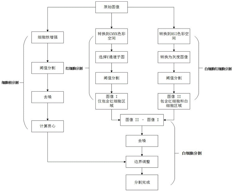
[0042] like figure 1 As shown, the specific implementation process of the white blood cell cytoplasm and nucleus segmentation method in the color blood cell image involved in the present invention is as follows:

[0043] In the original color blood cell image, the pixels in the nucleus area of ​​white blood cells have the largest contrast with pixels in other areas, and are the easiest to extract. In the present invention, the entire white blood cell area is first segmented, and then the nucleus part of the white blood cell is segmented. Finally, the nucleus part of the white blood cell is subtracted from the entire white blood cell area. Obtain the cytoplasmic fraction of the basal cells. In the present invention, white blood cell and red blood cell segmentation, red blood cell segmentation and cell nucleus segmentation can be performed simultaneously, whic...


PUM


Abstract

The invention discloses a method for partitioning cytoplasm and cell nucleuses of white blood cells in a color blood cell image. The method includes the following steps that the background region besides the white blood cells and red blood cells in the color blood cell image is removed, and a binary image I only including the region of the red blood cells and the region of the white blood cells is obtained; the white blood cells and the background region in the color blood cell image are removed, and a binary image II only including the region of the red blood cells is obtained; the binary image II is subtracted from the binary image I, and a binary image III only including the region of the white blood cells is obtained; the region of the cell nucleuses in the color blood cell image is enhanced, and a binary image IV only including the area of the cell nucleuses is obtained; the binary image IV is subtracted from the binary image III, and the area of the cytoplasm is obtained. The method has the advantages that the partitioning algorithm is simple in calculation, the partition of the white blood cells and the red blood cells and the partition of the red blood cells and the cell nucleuses can be conducted at the same time, the time expenditure is reduced, and partition errors are removed so that partitioning results are more accurate.

Description

technical field [0001] The invention belongs to the field of biomedical image processing, and in particular relates to a method for segmenting white blood cell cytoplasm and nucleus in a color blood cell image. Background technique [0002] There is an immune system in the human body, which is the most important defense system for the human body to resist the invasion of pathogenic bacteria, and is the third line of defense for the human body. It consists of immune organs, immune cells and immune molecules. Among them, white blood cells are the main component of immune cells, which can resist and eliminate invading pathogenic microorganisms by phagocytosis and antibody production. Leukocytes are a type of blood cells with nuclei. Normal people maintain a relatively stable level of leukocytes in the blood, about 4,000-10,000 / microliter. Any excess or deficiency of the normal level may be caused by a certain disease. [0003] Differential counting of white blood cells is one...

Claims


Application Information

Patent Timeline
IPC IPC(8): G06T7/00
Inventor 郑成云刘治张聪聪李晓梅刘晶肖晓燕
Owner SHANDONG UNIV
Learn More