Urine cell image classification and identification system and method

A technology of image recognition and urine cells, which is applied in the field of clinical medical technology applications, can solve problems such as poor scientific rigor, eye fatigue, misjudgment errors, etc., and achieve the effect of improving accuracy and reducing workload

Inactive Publication Date: 2013-04-03
TIANJIN UNIVERSITY OF TECHNOLOGY
View PDF2 Cites 6 Cited by
  • Summary
  • Abstract
  • Description
  • Claims
  • Application Information

AI Technical Summary

Problems solved by technology

However, the microscopic observation through the human eye not only easily fatigues the eyes of the observer, and then causes misjudgment errors, but also the subjective observation of the human eye is greatly affected by subjective factors, lacks objective evaluation standards, and has poor scientific rigor

Method used


Image

  • Urine cell image classification and identification system and method
  • Urine cell image classification and identification system and method
  • Urine cell image classification and identification system and method

Examples


Embodiment 1

[0018] Embodiment 1, urine cell image classification recognition system

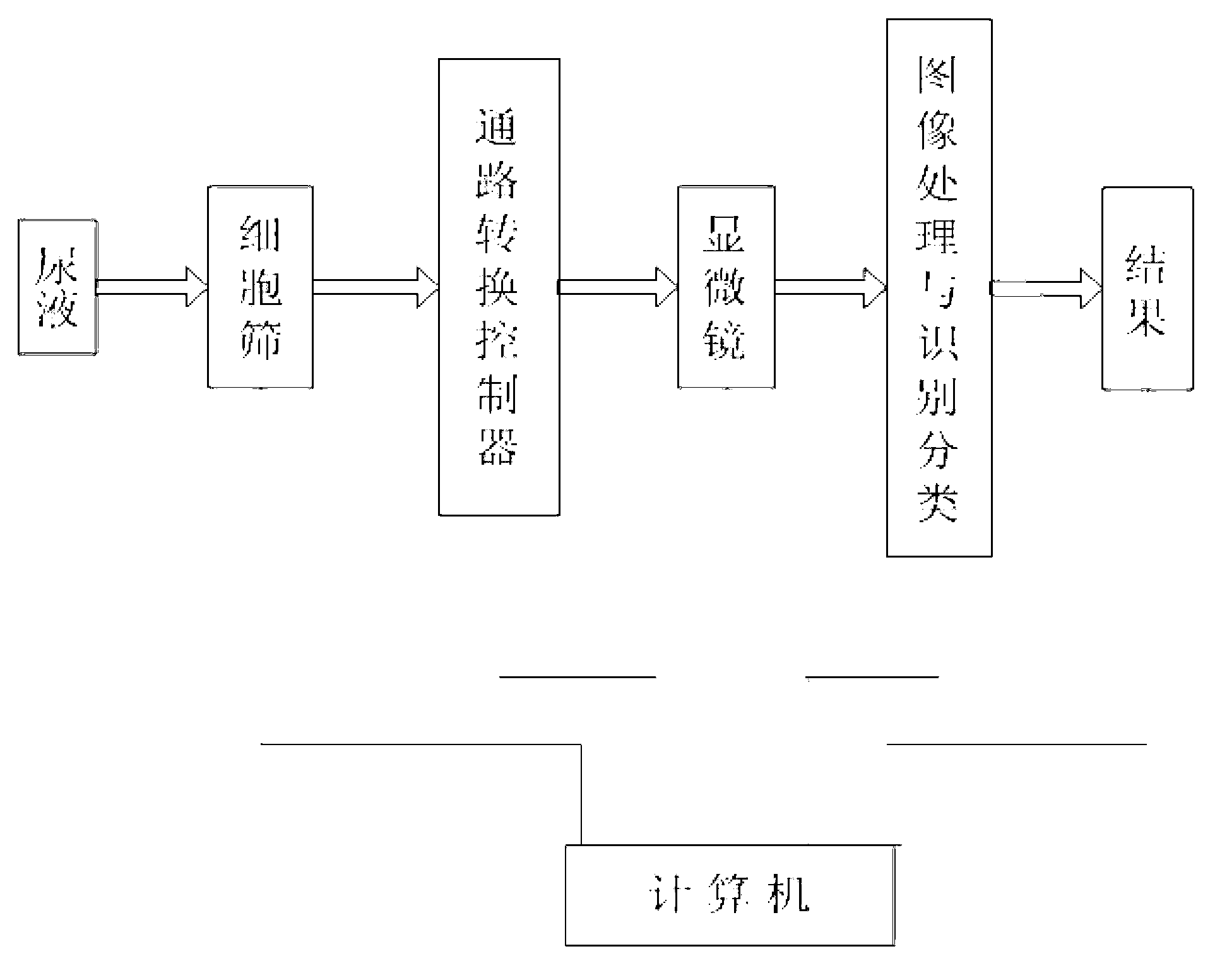
[0019] The urine cell image classification and recognition system consists of a cell screen, a path conversion controller, a microscope, an image recognition classifier and a computer;

[0020] cell sieve (eg figure 2 As shown), it is composed of molecular sieves of different specifications placed on the screening plate, specifically including type 3A, type 4A, type 5A, type 10X, and type 13X, and the pore diameters are about Then the urine to be tested is screened by the cells controlled by the rotation of the computer.

[0021] Path switching controller, its role is equivalent to a switch, the cell sieve screens out cells of different sizes, and controls the on and off through the computer, and transmits the cells of different sizes screened successively to the microscope in sequence;

[0022] The image recognition classifier adopts the support vector machine classifier to predict and classify all ...

Embodiment 2

[0023] Embodiment 2, the method for urine cell image classification and recognition

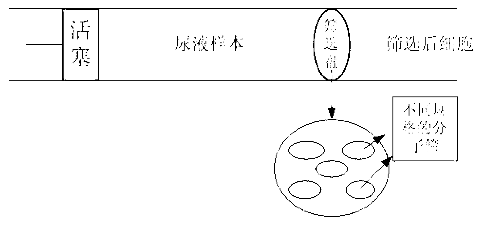
[0024] The specific steps of the urine cell image classification and recognition method provided by the present invention are as follows: figure 1 As shown, the method is a rapid and accurate method for the classification of urine occult blood cells, including:

[0025] The use of cell sieves for cell screening mainly uses physical methods for preliminary classification of urine samples. First, the urine cells are diluted, and then the pressure is provided through the piston in the pipeline to control the speed of the urine. Controlled by a computer, the rotating cell sieve Then the urine flows into the cell sieve at a speed of 0.1mm / s for screening, and then the rotating screening disc is controlled by the computer, and rotates to the next molecular sieve to increase the size of the screened cells Repeat this step to separate the cells of different sizes in the urine, and then the computer ...


PUM


Abstract

The invention relates to a urine cell image classification and identification system and a urine cell image classification and identification method. The urine cell image classification and identification system mainly comprises a cell sieve, a passage transformation controller, an image identification classifier and a computer. The urine cell image classification and identification method comprises the following steps of: firstly diluting urine cells, carrying out cell screening by using the cell sieve, rotating the screening tray inside the cell sieve to a molecular sieve with minimal specification, and controlling the screening tray to rotate to a next molecular sieve through the computer to achieve the requirement of enlarging the sizes of screened cells; repeating the step to separate the cells which are contained in urine and different in size, then controlling the on-off of the passage transformation controller through the computer to transmit the screened cells with different sizes to a microscope for image observation and acquisition, then transmitting to the computer, and then treating images to be detected through the image identification classifier by utilizing image preprocessing and separating technologies; and finally completing classification and identification.

Description

technical field [0001] The invention belongs to the technical application field of clinical medicine, and especially has important clinical significance for disease diagnosis, treatment monitoring and health general examination. Background technique [0002] In recent years, with the development of computer technology, digital image processing technology has developed rapidly, and has a wide range of application requirements in various fields such as aerospace, industrial production, resource environment, meteorology and traffic monitoring, culture and education, and has created a huge amount of Social value. In biomedical engineering, for example, the processing and recognition of microscopic images have been well applied in the automatic counting and structure automatic analysis of blood cells and the intelligent diagnosis of cells. With the aid of a microscope, people can observe the microscopic and ultramicroscopic structures of cells, and by analyzing their morphology,...

Claims


Application Information

Patent Timeline
Patent Type & Authority Applications(China)
IPC IPC(8): G01N21/84G06K9/62
Inventor 孙杰张静文秦颖博
Owner TIANJIN UNIVERSITY OF TECHNOLOGY
Why Eureka
Try Eureka