
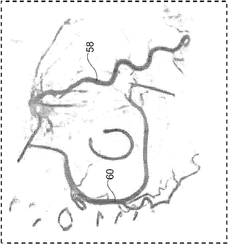
Visualization of the coronary artery tree

一种冠状动脉、动脉的技术,应用在重建冠状动脉的检查设备领域,达到快速且可靠诊断的效果

Active Publication Date: 2011-10-19
KONINK PHILIPS ELECTRONICS NV
View PDF3 Cites 1 Cited by
  • Summary
  • Abstract
  • Description
  • Claims
  • Application Information

AI Technical Summary

Problems solved by technology

However, it has been shown that this does not provide sufficient information to the user, i.e. for example a surgeon or a cardiologist
However, there may be a need to provide comprehensive information on current coronary status

Method used


Image

  • Visualization of the coronary artery tree
  • Visualization of the coronary artery tree
  • Visualization of the coronary artery tree

Examples


Embodiment Construction

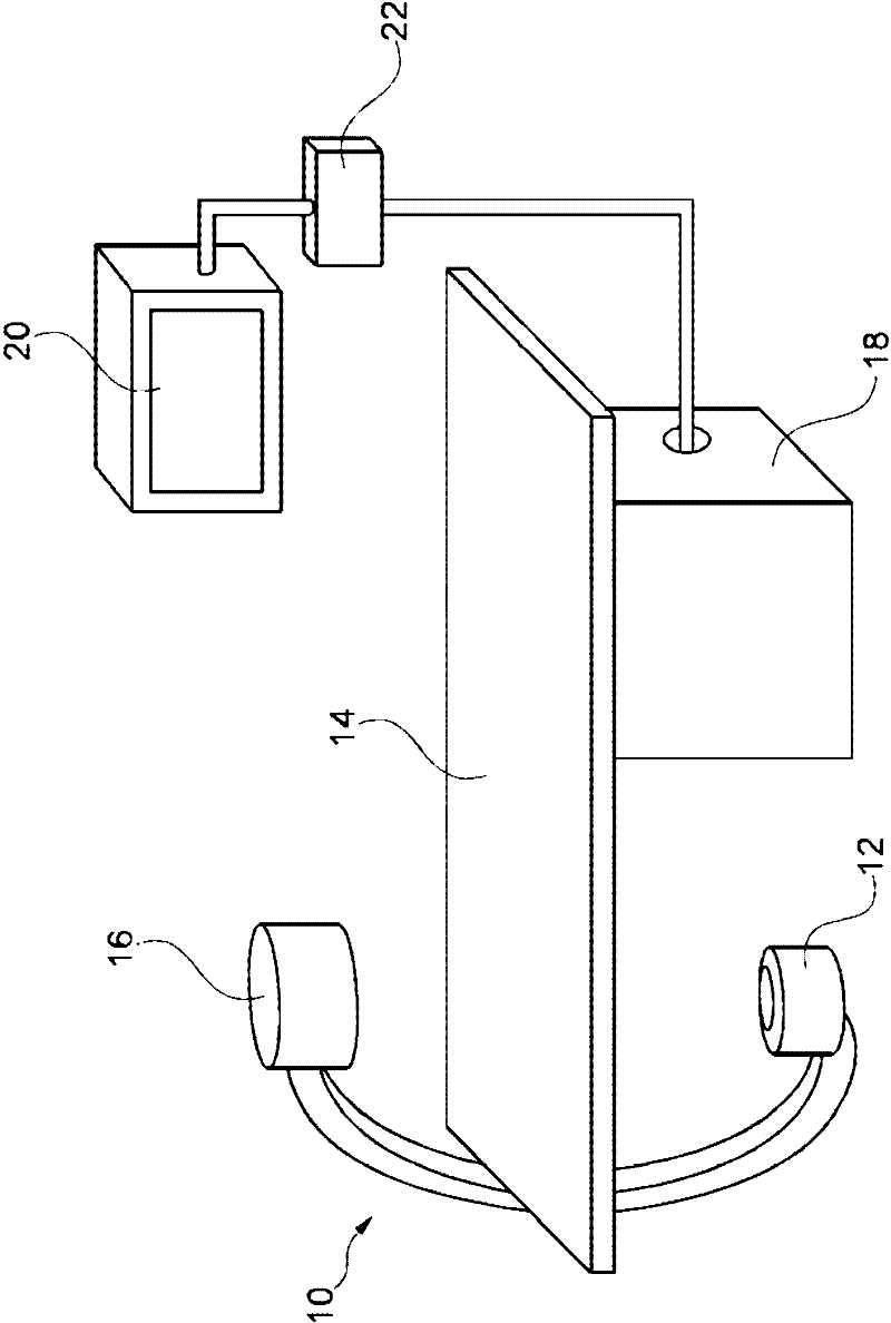
[0046] figure 1 An x-ray imaging system 10 with an examination device for reconstructing coronary arteries is schematically shown. The examination apparatus comprises an X-ray image acquisition device provided with an X-ray radiation source 12 for generating X-ray radiation. A station 14 is provided to receive subjects for examination. Furthermore, the X-ray image detection module 16 is located on the opposite side of the X-ray radiation source 12 , ie the subject is located between the X-ray radiation source 12 and the detection module 16 during the radiation process. The detection module 16 sends data to a data processing unit or calculation unit 18 which is connected to both the detection module 16 and the radiation source 12 . The computing unit 18 is located below the table 14 to save space in the examination room. Of course, it can also be located in different places, such as different rooms or laboratories. Furthermore, a display device 20 is arranged in the vicinit...


PUM


Abstract

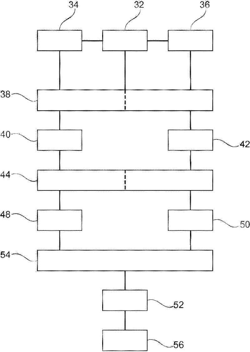
The present invention is related to a method for reconstruction of the coronary arteries and an examination apparatus for reconstruction of the coronary arteries. To provide improved coronary artery information, an apparatus and a method are provided where a gating signal is provided (32) and a first gated X-ray image sequence of one of the left or right branches of the coronary arteries is acquired (34) with injected contrast agent into the one of the left or right branches of the coronary arteries. Further, a second gated X-ray image sequence of the other branch of the coronary arteries is acquired (36) with injected contrast agent into said other branch. Then, a gated reconstructing (38) of the left and the right coronary artery is suggested and a volume data (40, 42) of the coronary arteries is generated. The volume data of the left and right coronary arteries is registered (44) in relation to time and space. Further; the registered volume data (48, 50) of the left and the right coronary arteries is combined and a combined coronary artery tree volume data set (52) is generated (54). Finally, the combined coronary tree volume data set is visualized (56).

Description

technical field [0001] The invention relates to a method for reconstructing a coronary artery and an examination device for reconstructing a coronary artery. Background technique [0002] During interventions, for diagnostic purposes—treatment planning or outcome control of interventions—it is known to visualize the left and right coronary arteries partially surrounding the heart by using so-called C-mode X-ray image generating devices. The visualization is generally based on the reconstruction of the coronary arteries with the aid of at least two acquired images. For acquisition, a contrast agent is selectively injected into the left or right branch of the coronary arteries (LCA or RCA). In other words, right branch image acquisition is separate from image acquisition for the left branch. Acquisitions are usually analyzed independently to make a diagnosis of the overall coronary tree. However, it has been shown that this does not provide sufficient information to the use...

Claims


Application Information

Patent Timeline
Patent Type & Authority Applications(China)
IPC IPC(8): A61B6/00G06T11/00
CPCA61B6/4441A61B6/466A61B6/481A61B6/541G06T11/006A61B6/504G06T2211/404
Inventor E·S·汉西斯M·格拉斯
Owner KONINK PHILIPS ELECTRONICS NV
Learn More