Three-dimensional ultrasonography-based early liver cancer diseased tissue target detection method

A three-dimensional ultrasound and early detection technology, applied in the field of imaging medicine, can solve problems such as complicated steps, low display accuracy, and human trauma, and achieve the effect of improving objectivity and accuracy, and enriching the amount of information

Inactive Publication Date: 2011-03-16
HUBEI JINGSHANG ENTERPRISE MANAGEMENT
View PDF0 Cites 10 Cited by
  • Summary
  • Abstract
  • Description
  • Claims
  • Application Information

AI Technical Summary

Problems solved by technology

In the past, the liver and other tissue structures were subjected to coarse needle puncture, forceps, incision, and resection, and then made into pathological sections, and then the target diagnosis was determined through a microscope. The steps were complicated and traumatic to the human body, and the display accuracy was low.

Method used


Image

  • Three-dimensional ultrasonography-based early liver cancer diseased tissue target detection method
  • Three-dimensional ultrasonography-based early liver cancer diseased tissue target detection method
  • Three-dimensional ultrasonography-based early liver cancer diseased tissue target detection method

Examples


Embodiment Construction

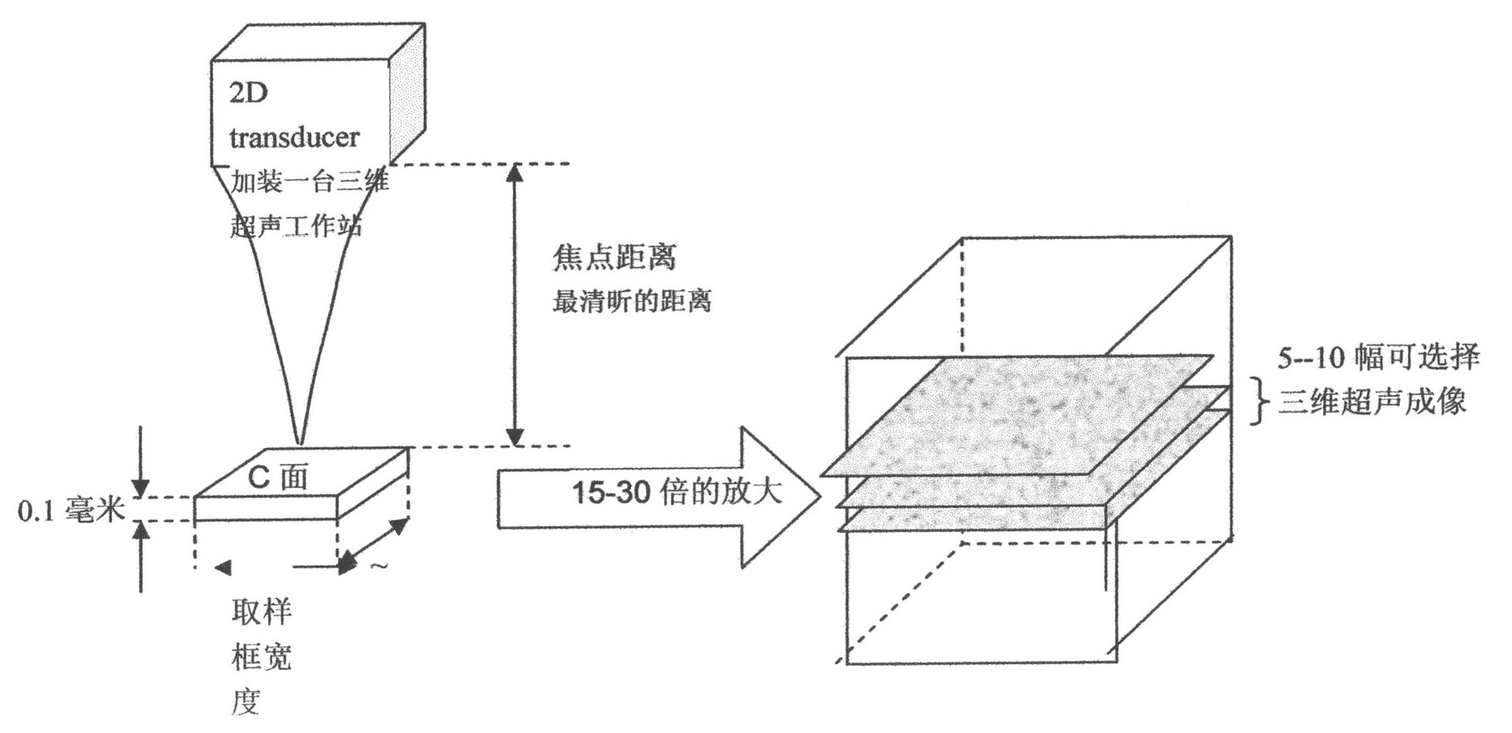
[0024] according to Figure 1-9 As shown, a method for early detection of liver cancer lesion tissue target by three-dimensional ultrasound imaging, the steps are as follows:

[0025] (1) Use the free arm to scan the three-dimensional ultrasound to reconstruct the C-plane;

[0026] (2) Use conventional B-ultrasound inspection first, then enter the three-dimensional inspection mode, and use the coordinate displacement method or sector scanning method to scan the target. The scanning line is in a specific mode, and the image within the reasonable minimum distance is formed. , apply the minimum three-dimensional reconstruction mode, and observe the C plane of the image;

[0027] (3) Observe the shape, brightness, distribution and internal structure of the target substantive image through appropriate image enlargement processing;

[0028] (4) Make these extremely valuable acoustic imaging information can be effectively reflected in a three-dimensional way;

[0029] (5) After st...


PUM


Abstract

The invention relates to a three-dimensional ultrasonography-based early liver cancer diseased tissue target detection method. The method comprises the following steps of: (1) scanning a three-dimensional ultrasonic reconstruction C surface by using a free arm; (2) scanning a target by a coordinate displacement method or a sector scanning method, and observing the C surface of an image in a minimum three-dimensional reconstruction mode; (3) amplifying and observing the shape, brightness, distribution condition and internal structure of an actual image of the target; (4) effectively reflecting the valuable ultrasonographic information in a three-dimensional mode; and (5) enabling the display precision of the image of the target to reach 0.1 millimeter-scale level sampling thickness. The three-dimensional ultrasonography-based early liver cancer diseased tissue target detection method has the advantages that: the imaging mode breaks through the limit of the action of an ultrasonic physical principle, a diseased tissue of which the minimum size is about 0.1mm can be observed, and an effect similar to a microscopic observation effect is achieved; a brand new image structure is generated to ensure that the information content of an ultrasonic image is greatly enriched and the objectivity and accuracy of image analysis are obviously improved; and the method lays a novel important basis for early diagnosis and treatment of various liver diseases.

Description

technical field [0001] The invention relates to the technical field of imaging medicine, in particular to a method for early detection of liver cancer lesion tissue targets by three-dimensional ultrasonic imaging. Background technique [0002] With the development of ultrasonic detection and imaging technology represented by digital technology, the various resolution capabilities and display characteristics of ultrasonic detection technology have made great progress, and more colorful audio-visual information can be obtained through the echoes of various tissues , but some extremely valuable sonographic information cannot be effectively reflected in general two-dimensional ultrasonography. [0003] At present, the detection of fetuses, hearts and other parts by three-dimensional ultrasound is all three-dimensional ultrasound applications for the purpose of stereoscopic observation. There is no method similar to microscopes to non-invasively make three-dimensional ultrasound ...

Claims


Application Information

Patent Timeline
Patent Type & Authority Applications(China)
IPC IPC(8): A61B8/08
Inventor 陈莉望鑫汪俊磊罗玲易秋萍
Owner HUBEI JINGSHANG ENTERPRISE MANAGEMENT
Why Eureka
Try Eureka