
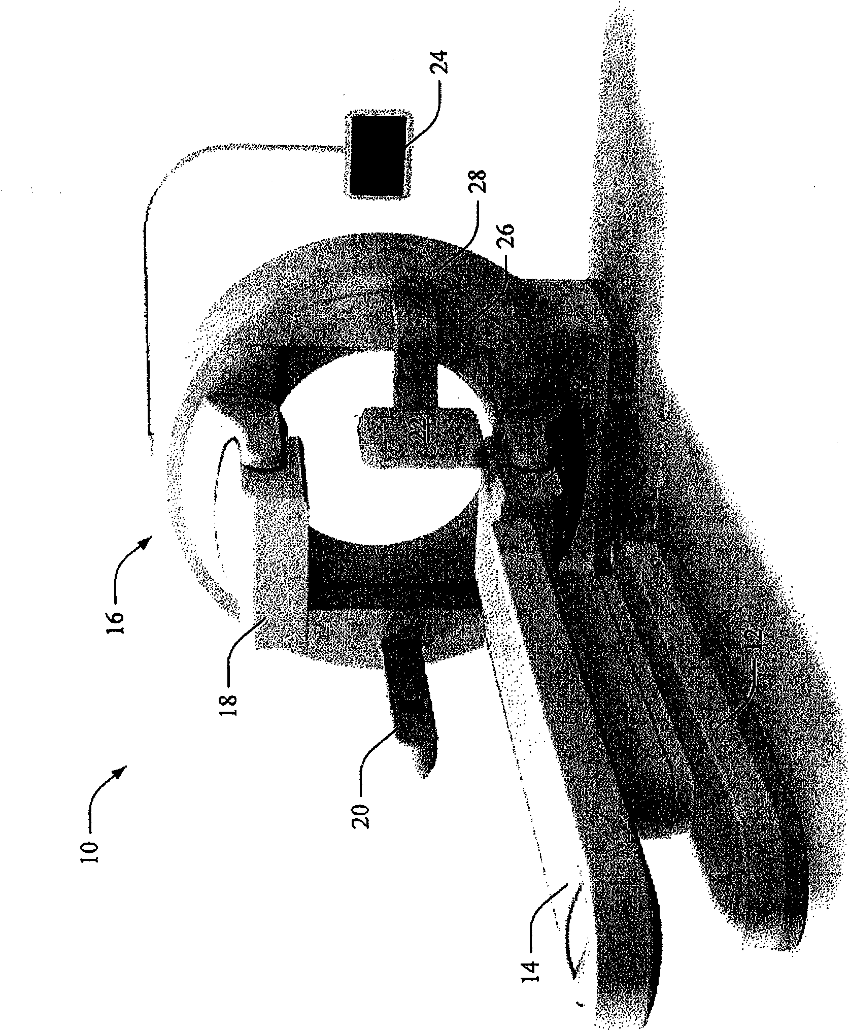
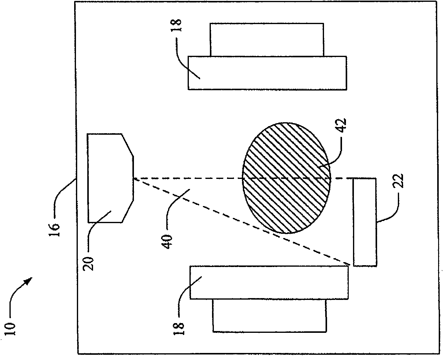
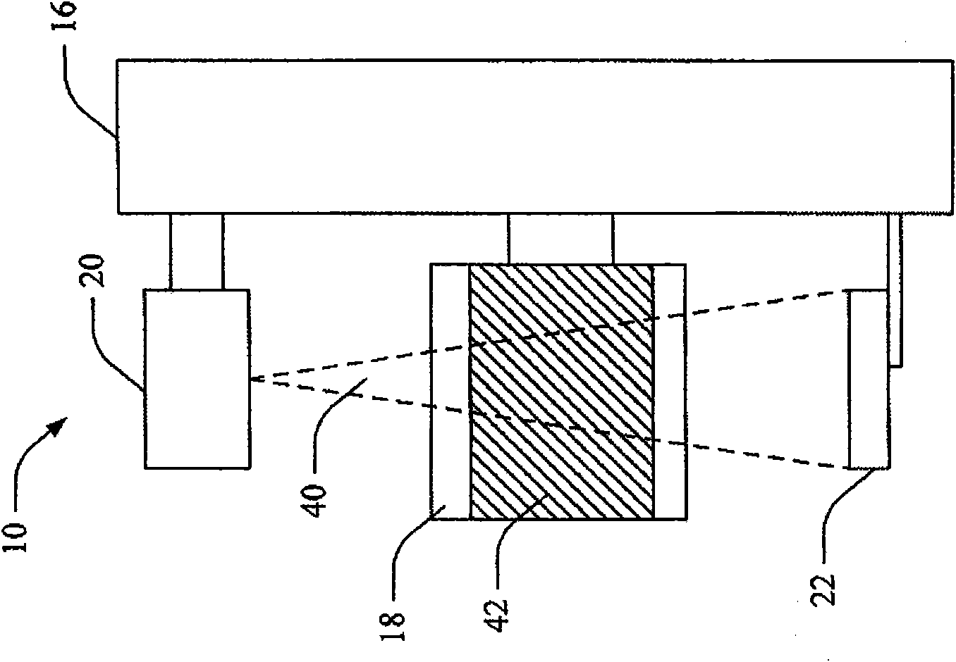
Nuclear medicine SPECT-CT machine with integrated asymmetric flat panel cone-beam CT and SPECT system

A technology for imaging systems and nuclear detectors, used in medical science, scientific instruments, radiation measurement, etc., and can solve problems such as X-ray component interference

Active Publication Date: 2014-08-06
KONINKLIJKE PHILIPS ELECTRONICS NV
View PDF3 Cites 0 Cited by
  • Summary
  • Abstract
  • Description
  • Claims
  • Application Information

AI Technical Summary

Problems solved by technology

Integrating the two systems into one gantry involves additional packaging constraints and SPECT detector motions that can interfere with X-ray components

Method used


Image

  • Nuclear medicine SPECT-CT machine with integrated asymmetric flat panel cone-beam CT and SPECT system
  • Nuclear medicine SPECT-CT machine with integrated asymmetric flat panel cone-beam CT and SPECT system
  • Nuclear medicine SPECT-CT machine with integrated asymmetric flat panel cone-beam CT and SPECT system

Examples


Embodiment Construction

[0038]The systems and methods described here involve combining a cone beam CT (CBCT) source with an offset flat panel detector and nuclear imaging head in a single patient imaging setup. With an offset flat panel detector, the size of the detector can be minimized compared to a full size CT detector, thereby occupying less space and allowing greater freedom of movement of the nuclear imaging head. Nuclear imaging provides physiological process and / or functional information that can be used for diagnosis, to assess the effect of treatment, etc. Adding another modality, such as co-registered CBCT, is useful in improving the reader's clinical confidence. In addition, CBCT information can be used to correct for attenuation of the emission data, improving the quantitative accuracy and quality of the images.

[0039] Other features involve locking mechanisms that ensure that the flat panel detector remains in place during CBCT scans and retracts out of the way when the CBCT source ...


PUM


Abstract

A volume cone beam CT scan is performed using a cone beam CTX ray source (20) and an offset flat panel x-ray detector (22) during nuclear (eg SPECT or PET) and CT scans of a patient. The field of view of the X-ray source overlaps the field of view of the two nuclear detector heads (18), and the offset of the X-ray detector (22) is such that there is no difference between the movement of the nuclear detector heads around the rotatable gantry (16). interference is minimized. Additionally, the locking mechanism (80) provides automatic locking of the X-ray detector (22) in each of the stowed and working positions, improving safety and CT image quality.

Description

technical field [0001] The present application finds particular application in diagnostic imaging systems, particularly cone beam computed tomography (CT) and single photon emission computed tomography (SPECT). However, it will be appreciated that the techniques may also be applied in other imaging systems, other medical situations or other medical techniques. Background technique [0002] Typical commercial system designs that combine both nuclear medicine (NM) and X-ray CT modalities, such as Philips Precedence, Philips Gemini, Siemens Symbia, etc., are configured in tandem geometry. In this configuration, a common patient table extends between two imaging field-of-view (FOV) regions axially separated by the space between the NM and the CT gantry. [0003] In other commercial system designs, such as in GE Hawkeye, the x-ray source and CT detector are mounted on a common gantry with NM detectors, but are still axially separated. Others have proposed the concept of using f...

Claims


Application Information

Patent Timeline
IPC IPC(8): A61B6/03G01T1/164
CPCA61B6/4435Y10T403/32254A61B6/4417A61B6/105A61B6/4476A61B6/4085A61B6/5241A61B6/4441A61B6/037A61B6/032A61B6/4291A61B6/4405A61B6/5229A61B6/4283
Inventor J·F·韦塞尔M·J·彼得里洛I·法默M·詹巴克施R·哈桑P·胡格J·J·斯特凡M·A·休伯
Owner KONINKLIJKE PHILIPS ELECTRONICS NV
Learn More