
Image processing device and ultrasonic diagnostic apparatus

Technology of an image processing device and ultrasonic diagnostic apparatus

Active Publication Date: 2011-01-26
SHENZHEN MINDRAY BIO MEDICAL ELECTRONICS CO LTD +1
View PDF0 Cites 0 Cited by
  • Summary
  • Abstract
  • Description
  • Claims
  • Application Information

AI Technical Summary

Problems solved by technology

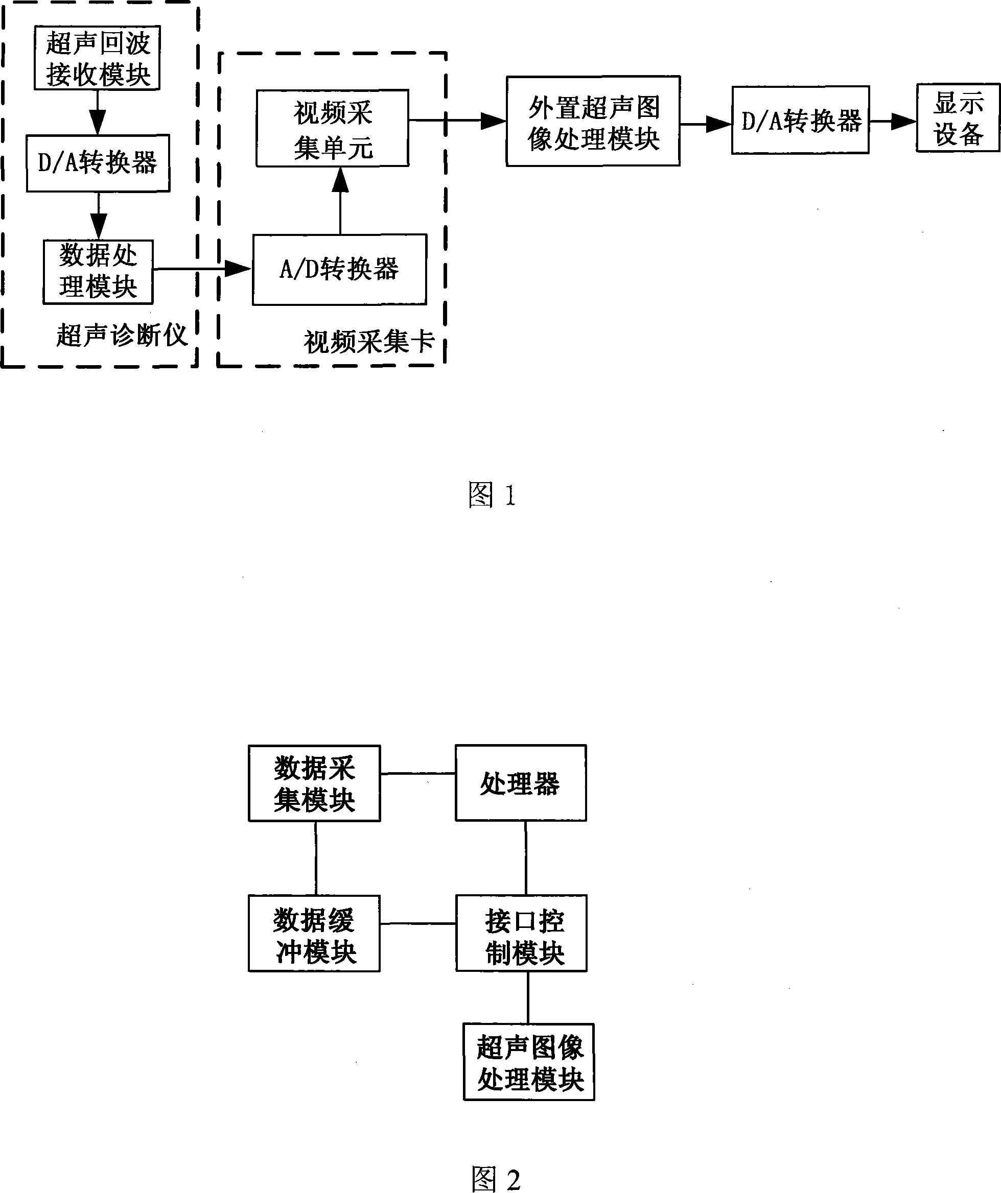
[0005] The existing technology uses an external ultrasonic image processing module to process the ultrasonic image again, and its main disadvantage is that it must rely on an external ultrasonic image processing module to complete the processing; and the prior art uses a video acquisition card for ultrasonic diagnosis When the video signal of the ultrasonic diagnostic instrument is collected as the data source for the reprocessing of the ultrasonic image, the image data of the ultrasonic diagnostic instrument has undergone a D / A conversion before being converted into a Video or S-Video signal output, and then in the video acquisition card. After an A / D conversion, the digital signal is transmitted to an external ultrasonic image processing module, such as a PC (Personal Computer, personal computer) for processing, and the processed data needs to undergo a D / A conversion again
Therefore, using an external ultrasonic image processing module for image processing requires one more D / A conversion and one A / D conversion compared to the ultrasonic diagnostic instrument, which will inevitably cause the loss of effective image data and directly affect the reprocessed image. Effect

Method used


Image

  • Image processing device and ultrasonic diagnostic apparatus
  • Image processing device and ultrasonic diagnostic apparatus
  • Image processing device and ultrasonic diagnostic apparatus

Examples


Embodiment 1

[0028] As shown in Figure 2, the image processing device includes a data acquisition module, a data buffer module, an interface control module, a processor, and an ultrasonic image processing module connected to the controller module; wherein, the data acquisition module adopts a CPLD chip, and the data buffer module adopts SSRAM, the memory that contains the stored program in the processor adopts the embedded processor.

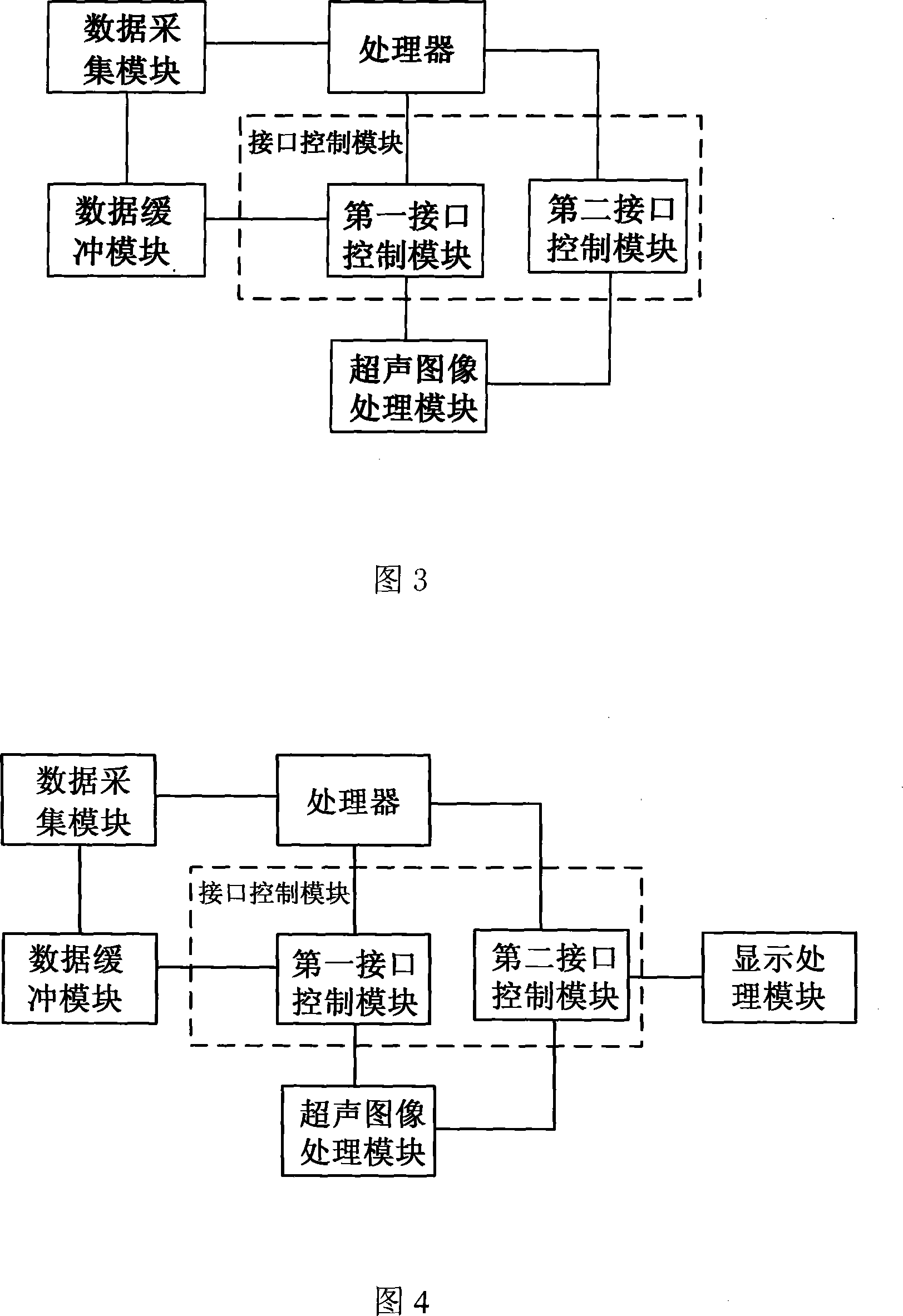
[0029] As shown in Figure 3, the interface control module includes a first interface control module and a second interface control module, wherein the first interface control module is connected with the processor, the data buffer module and the ultrasonic image processing module; the second interface control module It is connected with the processor and the ultrasonic image processing module.

[0030] The data acquisition module collects image data under the control of the processor, and the collected image data is stored in the data buffer module, and the ...

Embodiment 2

[0033] As shown in FIG. 4 , the difference between this embodiment and the first embodiment is that it further includes a display processing module connected to the second interface control module. The process flow of the image processing device for processing the ultrasonic image is shown in Figure 8, including the following steps:

[0034] (1) After power-on reset, the system loads relevant configuration files and the firmware (Firmware) of the embedded processor;

[0035] (2) After the loading is completed, the embedded processor starts to run the application program stored in the memory and enters the working state;

[0036](3) The embedded processor receives information such as the start acquisition command and the number of acquisition frames sent by the ultrasonic image processing module, and the embedded processor sets the acquisition control logic and transmits the original image data to the data buffer

[0037] (4) The ultrasonic image processing module queries the ...

Embodiment 3

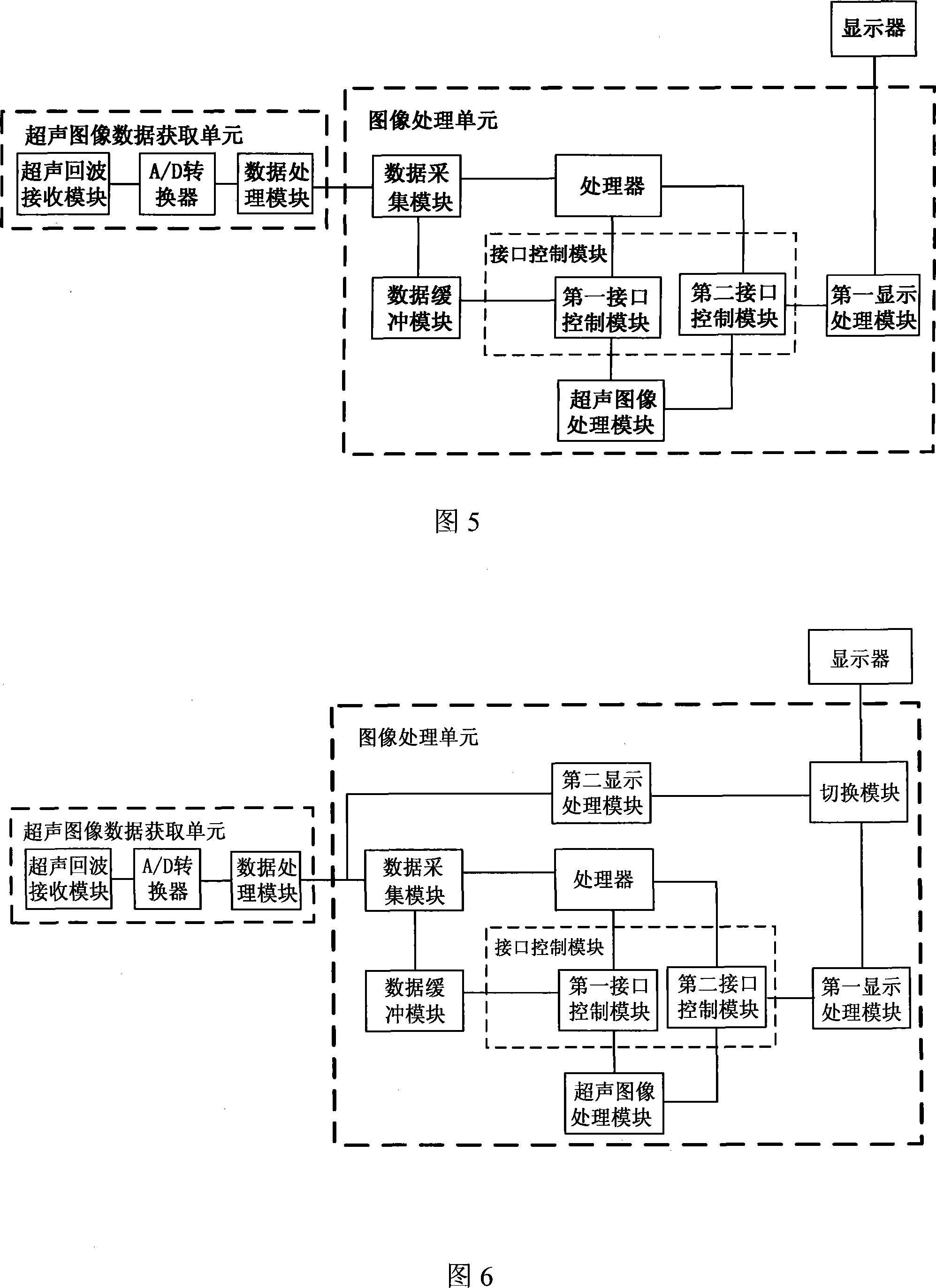
[0043] As shown in Figure 5, the ultrasonic diagnostic apparatus of this example includes an ultrasonic image data acquisition unit and an image processing unit, and the two are connected in sequence; wherein, the image processing unit includes a data acquisition module, a data buffer module, an interface control module, a processor, an ultrasonic The image processing module and the first display processing module; the processor is respectively connected with the data acquisition module and the interface control module; the data buffer module is respectively connected with the data acquisition module and the interface control module; the ultrasonic image processing module is connected with the interface control module.

[0044] Wherein, the ultrasonic image data acquisition unit includes an ultrasonic echo receiving module, an A / D converter and a data processing module connected in sequence; the interface control module includes a first interface control module and a second inte...


PUM


Abstract

The invention discloses an image processing apparatus and an ultrasonic diagnostic device, the image processing apparatus comprises a data acquisition module, a data buffering module, an interface control module, a processor and an ultrasonic image processing module; the data acquisition module acquires image data under control of the processor and stores the acquired image data into the data buffering module, the image data in the data buffering module is transmitted to the ultrasonic image processing module by the interface control module to be processed, the processed image data can be output by the interface control module. The image processing apparatus provided by the invention can perform three-dimensional imaging and wide radial imaging or the like for the ultrasonic image data; can avoid loss of the effective image data when the ultrasonic diagnostic device uses an external image processing module, can ensure the image effect after processing the ultrasonic image again, thereby improving the accuracies of the detection and the analysis by using the ultrasonic diagnostic device.

Description

technical field [0001] The invention relates to image processing and medical equipment, in particular to an image processing device and an ultrasonic diagnostic instrument. Background technique [0002] Ultrasound imaging has the advantages of non-invasive, non-destructive, and non-ionizing radiation, and has been paid more and more attention by people. On the basis of the direct imaging of the ultrasonic diagnostic instrument, it is often necessary to reprocess the ultrasonic imaging data to achieve more advanced functions, such as three-dimensional imaging, wide-diameter imaging and other functions. The current ultrasonic diagnostic instrument itself does not have the function of ultrasonic image reprocessing, and must rely on an external ultrasonic data processing module. This data processing module is completely separated from the ultrasonic diagnostic instrument in structure and works independently of the ultrasonic diagnostic instrument. [0003] At present, the gener...

Claims


Application Information

Patent Timeline
Patent Type & Authority Patents(China)
IPC IPC(8): A61B8/00
Inventor 王哲刘阳春何绪金
Owner SHENZHEN MINDRAY BIO MEDICAL ELECTRONICS CO LTD
Learn More