
Endoscopic image processing apparatus

An image processing device and endoscope technology, applied in the direction of image data processing, image data processing, endoscope, etc., can solve the problems of not having, not having lesion missed, and electronic endoscope system not having, etc. Achieve the effect of improving observation efficiency

Active Publication Date: 2008-12-10
OLYMPUS CORP
View PDF1 Cites 20 Cited by
  • Summary
  • Abstract
  • Description
  • Claims
  • Application Information

AI Technical Summary

Problems solved by technology

[0007] On the other hand, the electronic endoscope system of Patent Document 1 has a structure for associating the position information of the distal end of the endoscope when the image is captured with the image, but does not have a structure that can prevent the For example, there is no structure capable of displaying information about the lesion confirmed when the endoscope is inserted when the endoscope is pulled out.
In other words, the electronic endoscope system of Patent Document 1 does not have a structure capable of solving the aforementioned problems arising in the aforementioned observation method

Method used


Image

  • Endoscopic image processing apparatus
  • Endoscopic image processing apparatus
  • Endoscopic image processing apparatus

Examples


Embodiment Construction

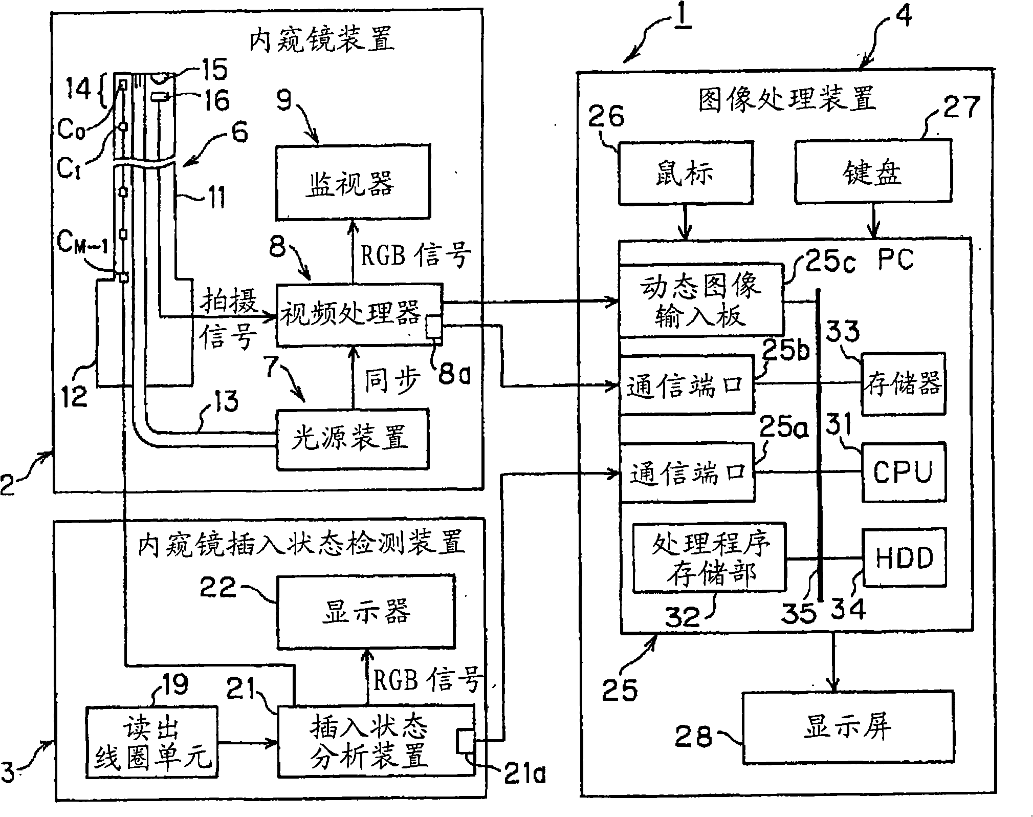
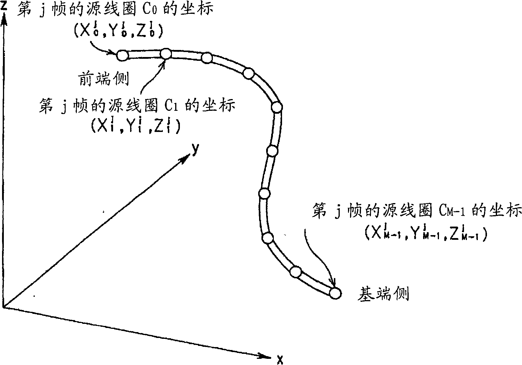
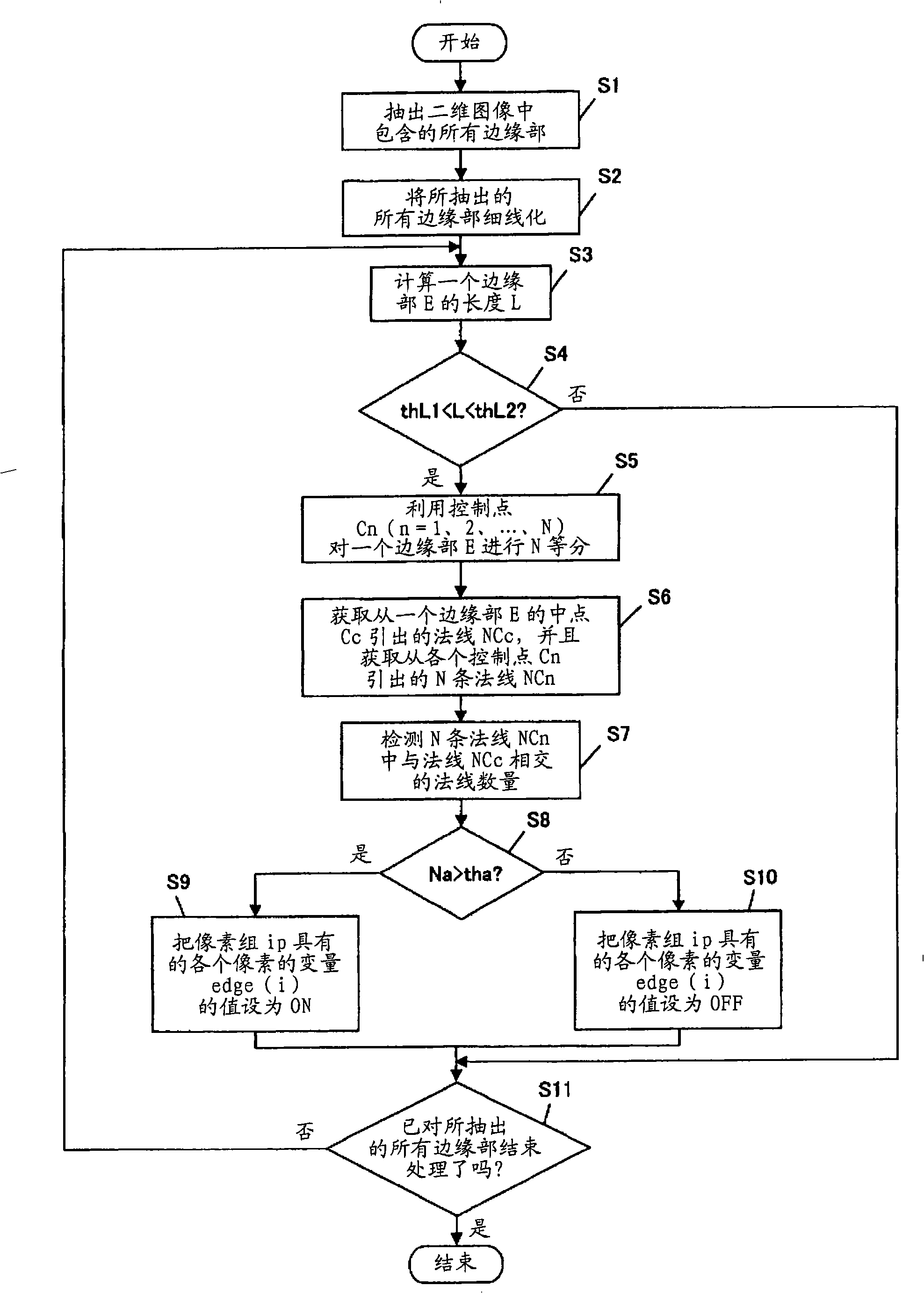
[0031] Hereinafter, embodiments of the present invention will be described with reference to the drawings. Figure 1 to Figure 9 It relates to an embodiment of the present invention. figure 1 It is a diagram showing an example of a configuration of a main part of a living body observation system using an image processing device according to an embodiment of the present invention. figure 2 is expressed in figure 1 The endoscope insertion state detection device is detected in the figure 1 A diagram of the coordinates of the source coil in the insertion section of the endoscope. image 3 means that when a lesion with a raised shape is detected, figure 1 A flowchart of a part of the processing performed by the image processing device. Figure 4 means that when a lesion with a raised shape is detected, figure 1 The image processing device followed by image 3 A flowchart of the processing performed for the processing. Figure 5 is to usefigure 1 A diagram of an example of a...


PUM


Abstract

The invention provides an endoscopic image processing apparatus capable of improving the observing efficiency for using endoscopic compared with the previous one, including: an image acquiring unit which acquires images according to subject images picked up over time by an endoscope inserted into an object to be examined; a lesion detecting unit which detects a lesion in an image each time the image is acquired; a display unit which displays the images; and an image display control unit which controls display conditions of a plurality of images including at least a lesion image in which a lesion has been detected by the lesion detecting unit out of the images acquired by the image acquiring unit.

Description

technical field [0001] The present invention relates to an endoscope image processing device, and more particularly, to endoscope image processing for controlling the display state of an image corresponding to an image of a subject captured by an endoscope inserted into a subject with the passage of time device. Background technique [0002] An endoscope system including an endoscope and the like has been widely used in industrial fields, medical fields, and the like. In particular, endoscope systems in the medical field are mainly used for purposes such as observation of various organs in a living body. Furthermore, as an endoscope system used for the aforementioned application, there is an electronic endoscope system proposed in Patent Document 1, for example. [0003] The electronic endoscope system of Patent Document 1 is configured to include: an imaging unit that uses an imaging element disposed at the front end of the endoscope to take pictures of the inside of the ...

Claims


Application Information

Patent Timeline
Patent Type & Authority Applications(China)
IPC IPC(8): A61B1/00A61B1/04G02B23/24G06T1/00G16Z99/00
CPCA61B1/0005A61B1/05G06T7/0012G06T2207/10068G06T2207/30028A61B2034/102A61B90/36A61B34/20A61B90/361A61B2034/105A61B2034/2051G16H40/63G16Z99/00
Inventor 西村博一
Owner OLYMPUS CORP
Learn More