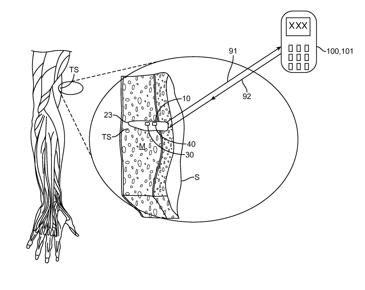
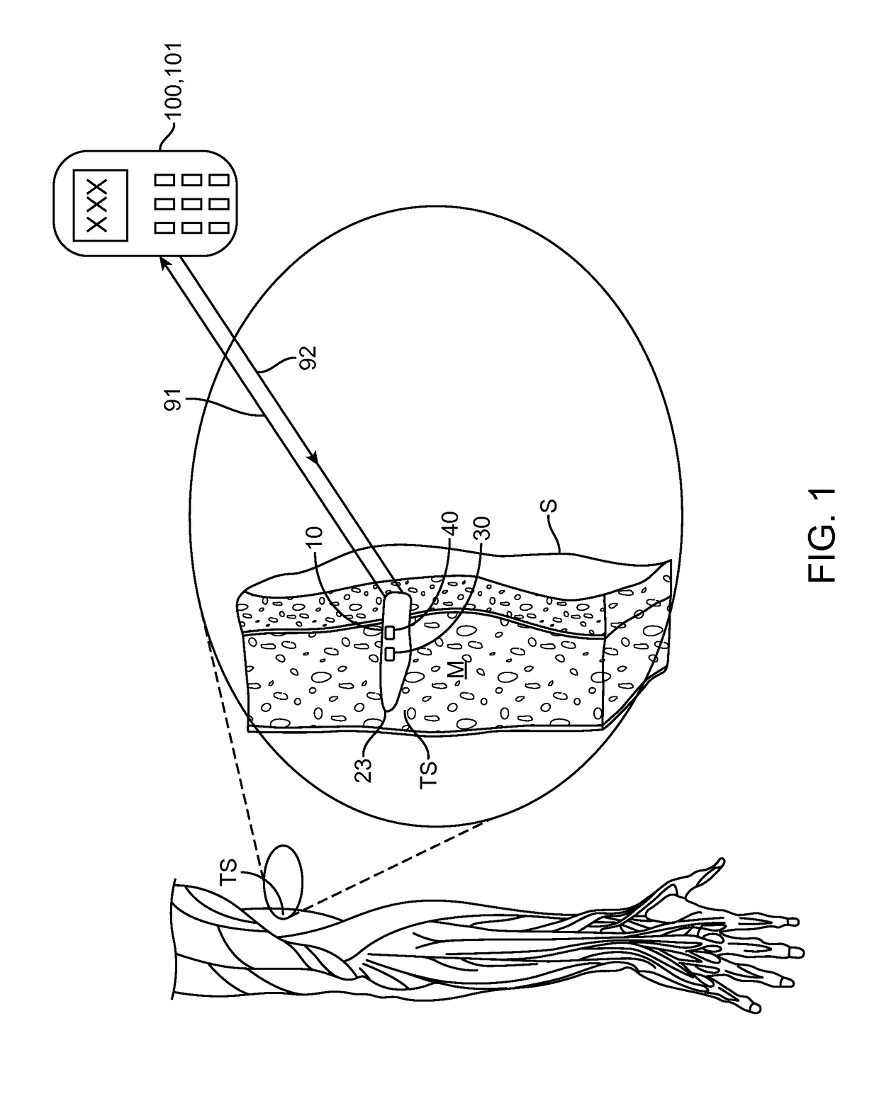
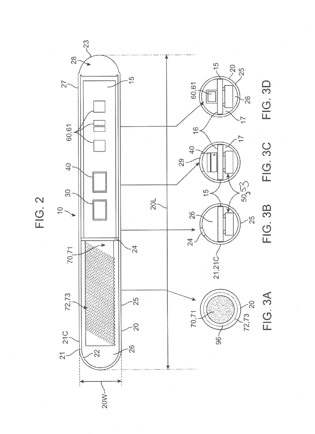
[0009]In various embodiments of the invention, the apparatus may include a magnetic hook which may comprise a magnet or a non-magnetic ferrous structure positioned on or within the housing to allow the apparatus to be removed from the tissue site or otherwise manipulated beneath the skin to facilitate removal using a removal tool containing a magnet or magnetized portion. The force of magnetic attraction between the hook and the removal tool magnetic portion being sufficient to pull the apparatus from the tissue site to the skin surface (i.e., beneath the skin surface) so that the apparatus can be easily removed with a very shallow incision in the skin. The removal tool may comprise a magnet itself or a finger grippable shaft attached to the magnet, allowing a physician to have finer control over the second magnet. In a particular embodiment, the magnet on the removal tool may comprise an adjustable electromagnet allowing the physician to adjust the amount of magnetic force used to remove the apparatus. In a related embodiment, the magnetic hook may comprise a ferrous-based suture or wire, herein, “a pull wire”, attached to the exterior surface of the housing which allows the physician to first pull the wire or suture to the skin surface using the removal tool and then pull the apparatus out to the tissue using the pull wire. In some embodiments, the pull wire can include a loop on its non-attached end to facilitate grasping of the wire once it is brought near the skin surface. The benefits of using the pull wire include one or more of the following: i) less magnetic force is required to remove the pull wire then that required to remove the whole housing allowing use of a less powerful second magnet; and ii) the physician can more easily and controllably remove the entire apparatus using the pull wire then removal by magnetic attraction alone which should reduce both tissue trauma and pain. In some embodiments, a combination of an internal magnetic hook and an external ferrous-based pull wire can be used to further facilitate atraumatic removal of the apparatus. Removal of the apparatus can also be facilitated by fabricating portions of the housing from radio-opaque, echogenic or other materials visible under a selected medical imaging modality and / or attaching to the housing one or more medical imaging markers such as radio-opaque and / or echogenic markers.
[0010]In various embodiments of the invention, all or a portion of the apparatus housing can be fabricated from shape memory materials such as nickel titanium alloys allowing the apparatus to be placed at the selected tissue sit; with the apparatus having a first size and shape and then upon exposure to body temperature within the tissue site, the apparatus expands to a second size and shape. In particular embodiments, the housing can be configured expand so as to anchor the housing at the tissue site. For example, in one or more embodiments, the housing can be fabricated from shape memory materials configured to expand to an hour glass or other like shape with the ends of the housing flaring out to hold the apparatus in place at the tissue site. In use, such embodiments allow the housing to have a smaller size to facilitate placement at the selected tissue site and then to expand to the larger size and / or shape to anchor the apparatus at the tissue site so as minimize movement of the apparatus once placed at the site. In related embodiments, all or a portion of the housing can be fabricated from shape memory materials having pseudo-elastic properties such as nickel titanium alloys (an example including NITINOL) configured to undergo a change in stiffness upon exposure to body temperature. Typically, this change will entail going from a more rigid state to a more flexible state (though the reverse is also contemplated). Such embodiments allow the apparatus to have a more rigid quality to facilitate insertion at the tissue site and then once placed, become more flexible to allow the housing to bend and flex with movement of the body including movement of tissue at the tissue site. In one embodiment, the center portion of the housing can be configured from a shape memory material which becomes elastic at body temperature allowing the center portion of the housing to bend and flex with movement of body tissue. Such embodiments of the housing having pseudo elastic properties also facilitate removal of the apparatus from the patient's body and reduce trauma since the housing can bend when grasp by a forceps or attracted by a magnetic removal tool allowing the housing to be bent up toward the skin surface or otherwise manipulated while imparting less force to surrounding tissue.
[0011]In other aspects of the invention, embodiments of the measurement apparatus can be adapted for numerous applications in the medical field including diagnostic and prognostic applications. Such applications can include, for example, tumor monitoring including monitoring a tumor for the efficacy of chemotherapy by using the oxygenated state of tissue in and around the tumor as an indication of the size and viability of the tumor. In use, such tumor monitoring applications allow a course of chemotherapy to be titrated responsive to the effects on the tumor of the chemotherapy. This improves both treatment efficacy and long term patient tolerance. For example, the dose of a particular chemotherapeutic compound with side effects such as nausea could be reduced upon determination that the tumor is shrinking, improving patient tolerance.
[0012]In another application, an embodiment of the oxygen saturation measurement apparatus can be adapted for monitoring the health and / or viability of an organ transplant by measuring the oxygenated state of tissue in and around the organ. Change such as decreases in oxygen saturation including rapid decreases being indicative of tissue rejection or the onset of tissue rejection. This in turn, allows time for rapid medical intervention to save the transplanted organ. In another application, embodiments of the apparatus can be used to monitor for oxygen saturation in the extremities of diabetic patients (who are prone to develop neuropathy in their extremities) allowing for medical intervention before the development of neuropathy. In still other applications, embodiments of the invention can be used for monitoring patients who have chronic obstructive pulmonary disease (COPD) or other respiratory disorders allowing for early diagnosis and treatment of an adverse respiratory state in such patients. Related embodiments can be use to monitor oxygen saturation levels of patients having congestive heart failure (CHF) again allowing for early diagnosis and treatment of various related conditions, e.g., pulmonary edema, before they become life threatening. In still other applications, embodiments of the invention can be used to monitor the progress of wound healing at a selected tissue site allowing for the early detection and treatment of acute conditions such as infection as well as monitoring the longer term progress of the wound healing process. Infection may be characterized by either a rise or fall in tissue oxygen saturation at the site depending upon the type of bacteria. For example, changes (e.g., lower) in the levels of oxygenation may be utilized as biomarkers / predictors of slower or other atypical healing requiring medical intervention (e.g., through the administration of one or more drugs, such as growth hormone, erythropoietin or other like compounds). Such predictions can be achieved by developing correlations between the time course of the healing process and levels of tissue oxygen saturation at site including rates of change of oxygen saturation. Still other related applications are also contemplated by various embodiments of the invention as will be known to those skilled in the medical diagnostic and other related arts. The features and attributes of the oxygen saturation measurement apparatus can be customized for each application, with such features including one or more of the size, shape, surface coatings, emitted and detector configuration and the wavelength, intensity and duty cycle of the emitted wavelength and other like factors. For example, the wavelength, intensity and duty cycle of the emitted light may be fine tuned for increased sensitivity to detect lower levels of blood oxygen saturation found in one or more of deep vein thrombosis, infected or tumerous tissue.


