
Implants for inducing soft and hard tissue integration

a soft and hard tissue integration and implant technology, applied in the field of biocompatible implants, can solve the problems of biomolecules becoming biologically inactive, low stability of biomolecule fixation, enhanced immunoreactivity, etc., and achieve good cell spreading, decreased inflammation, and no toxic

Active Publication Date: 2017-12-26
NUMAT BIOMEDICAL
View PDF9 Cites 1 Cited by
  • Summary
  • Abstract
  • Description
  • Claims
  • Application Information

AI Technical Summary

Benefits of technology

[0015]Surprisingly, the present invention provides an implant which overcomes the technical disadvantages of implants previously exposed. Accordingly, the present inventors have now surprisingly found that by binding a flavonoid or a methoxytryptophol, covalently or physically adsorbed, an ester thereof, and / or a pharmaceutically acceptable salt thereof and / or a combination thereof to a metal, metal alloy or metal oxide surface, such as a titanium and / or a titanium alloy surface, of a biocompatible implant, the osseointegrating and soft tissue attachment properties of the implant may be greatly facilitated. Such a biocompatible implant has not previously been known nor implied within the field.
[0016]The implant of the present invention presents an improvement of the attachment of the active biomolecule, in this case antioxidants, to the surface of the metal base. Furthermore, this attachment is improved when a linker with certain characteristics is used, and even more when a reduction of the complex linker-antioxidant is performed. This technical effect is demonstrated by showing that in implants wherein the antioxidant has been attached by covalent binding to the implant surface, the antioxidant is not released to the media (see examples 1, 2 and 3).
[0017]Furthermore, both human mesenchymal stem cells and gingival fibroblasts cultured on implants where the antioxidant has been covalently attached showed superior differentiation than cells cultured on implants where the antioxidant was simply physically attached. This surprising effect has been demonstrated by showing that genes involved in extracellular matrix production and organization and in regulation of cell adhesion, such as Collagens and Runx2, increased their expression in cells cultured on implants where the antioxidant has been covalently attached (see examples 2 and 3). This ability makes the implant of the present invention an ideal alternative in treatments where regeneration of both hard and soft tissues is required, such as in dental implants for the screws and abutments.
[0018]Additionally, the implant of the present invention is able to minimize undesired physiological effects occurred normally around the implanted zones such as inflammation. In cells cultured on implants with antioxidants covalently attached, the expression of interleukin-6 mRNA levels decreased as showed in example 2.

Problems solved by technology

Such binding has often been carried out using for example chemical reactants such as formalin or glutaraldehyde, but the reactive nature of these agents often leads to the biomolecules becoming biologically inactive and / or with enhanced immunoreactivity, which is of course undesirable.
Drawbacks are the rather low stability of biomolecule fixation, a non-defined release behaviour of biomolecules and possible conformational changes of the directly adsorbed molecules.
However, if more than one biomolecule is considered for immobilization, it is difficult to combine different molecules in a defined way on the surface.
Moreover, methoxytryptophols have never been reported in the context of bone regeneration or implants and surface coatings for improved osseointegration and soft tissue attachment.
However, there is no mention of the use of flavonoids for improved osseointegration or soft tissue attachment.
However, the application describes the use of flavonoids in bone graft applications with demineralized bone matrix and collagen sponges, and does not describe the use of flavonoids covalently attached to implant surfaces.
Furthermore, there is a limitation of most of the available techniques with regards to the physical absorption (labile union) of these compounds onto the surface of the implant.

Method used


Image

  • Implants for inducing soft and hard tissue integration
  • Implants for inducing soft and hard tissue integration
  • Implants for inducing soft and hard tissue integration

Examples


example 1

Conclusions Example 1

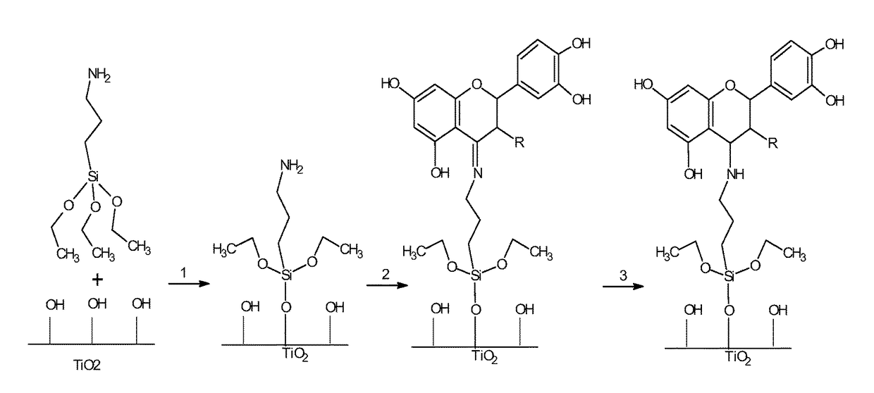
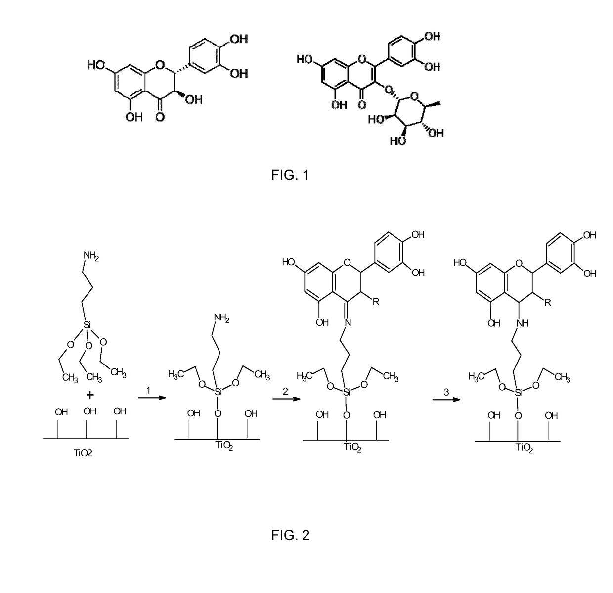
[0091]Flavonoids taxifolin and quercitrin can be grafted to Ti surfaces either by adsorption, drop casting and covalent linking methods. UV irradiation and passivation are effective pretreatments for Ti surface activation before the immobilization of the biomolecule.

[0092]Flavonoid-coated samples by adsorption and drop casting showed a high release of the flavonoid from the surface after 24 h incubation in water at physiological conditions.

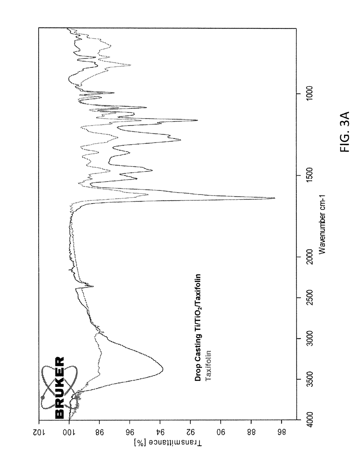
[0093]Covalently grafted samples showed different release behaviors depending on the reduction step. Non-reduced samples showed a low release of the flavonoid from the surface in water after 24 h incubation, while reduced samples using the same conditions did not show release of the flavonoid. As FTIR-ATR analysis of covalently immobilized samples showed the presence of the flavonoids on the Ti surface both for non-reduced and reduced samples, this demonstrates that the flavonoids are covalently linked to the substrate through...

example 2

Conclusions Example 2

[0119]None of the flavonoid-coated surfaces were toxic for HGF. After 24 hours of culture on the different surfaces, HGF possessed the typical fibroblastic spindle-shaped morphology and were distributed throughout the entire surface, without differences among the groups. This demonstrates that despite of the chemical modifications of the metallic surface of the implant by binding the linker and the flavonoid, toxicity has not been increased. Cytoskeleton and nuclei staining also revealed good cell spreading on all surfaces and the formation of prolongations and filopodia to contact each other and to attach to surfaces.

[0120]The different surface modification methods were effective in decreasing inflammation in HGF cells, as shown by the IL-6 mRNA levels. However, only flavonoids that were covalently attached to the implant surface were effective increasing the differentiation of HGF, as shown by the COL3A1 mRNA levels. Thus, to regenerate the gingival soft tissu...

example 3

Conclusions Example 3

[0137]None of the surfaces was toxic for hUC-MSCs. After 24 hours of culture on the different surfaces, hUC-MSCs showed the typical morphology with long thin cell bodies and prominent nucleous, which were placed throughout the entire surface, without differences among the groups.

[0138]As regards to the differentiation profile of the hUC-MSCs, cells cultured on implants that had flavonoids covalently attached showed superior differentiation than cells cultured on flavonoid drop-casted implants, as shown by the higher collagen-1, osteocalcin and, more importantly, runx2. Runx2 is a master organizer of gene transcription in developing and maturing osteoblasts, which are the main cells in hard tissue supporting osseointegrated implants.


PUM

PropertyMeasurementUnit
pHaaaaaaaaaa
diameteraaaaaaaaaa
diameteraaaaaaaaaa

Abstract

The present invention provides a biocompatible implant comprising one or more metal(s), metal alloy(s), metal oxide(s) or a combination thereof, wherein an antioxidant compound selected from the group of flavonoids or methoxytryptophols, an ester thereof, a pharmaceutically acceptable salt thereof or a combination thereof, is / are coated to at least a part of a metal, metal alloy or metal oxide surface of said biocompatible implant. This implant is useful for replace bone tissue in vertebrate animals, and furthermore restore the normal function of said tissue, mainly due to its ability of induce osseointegration and soft tissue attachment.

Description

FIELD OF THE INVENTION[0001]The present invention relates to the field of biocompatible implants, in particular biocompatible implants comprising at least one metal such as titanium, zirconium, tantalum, hafnium, niobium, chromium-vanadium alloy and stainless steel, or an alloy thereof. More specifically, the present invention relates to biocompatible implants comprising a metal, metal alloy and / or metal oxide surface which comprise a coating providing for improved osseointegration and soft tissue attachment of said biocompatible implant when implanted into a mammalian body.BACKGROUND OF THE INVENTION[0002]The use of implants to restore the function of traumatized or degenerated connective tissues and thus to improve the quality of life of a patient has become widespread. The long-term success of dental implants largely depends on rapid healing with safe integration into the jaw bone and soft tissue. The integration of the dental implant intra-osseus component and the transmucosal c...

Claims


Application Information

Patent Timeline
Patent Type & Authority Patents(United States)
IPC IPC(8): A61L27/06A61L27/28A61L27/04A61L27/54A61L31/16A61L31/08A61L31/02
CPCA61L27/06A61L27/04A61L27/28A61L27/54A61L31/08A61L31/16A61L31/022A61L2430/12A61L2400/18A61L2430/02
Inventor MONJO CABRER, MARTARAMIS MOREY, JOANA MARIACORDOBA INSENSE, ALBASATUE SAH N, MARIAGOMEZ FLORIT, MANUEL
Owner NUMAT BIOMEDICAL
Learn More