
Method of imaging localized infections

a localized infection and imaging technology, applied in the field of localized infection imaging, can solve the problems of inability to identify the location and/or the extent of infection in the site of localized infection, the infection can damage the host tissue, and the conventional method of diagnosis cannot solve the problem of chronic infection,

Inactive Publication Date: 2015-02-24
PURDUE RES FOUND INC
View PDF133 Cites 2 Cited by
  • Summary
  • Abstract
  • Description
  • Claims
  • Application Information

AI Technical Summary

Benefits of technology

[0004]An inflammatory response is one of the first responses to infection. Inflammation results in increased blood flow to the area of infection, release of chemicals (e.g., chemoattractants) to attract leukocytes to the site of infection, and migration of immune cells, such as monocytes and macrophages, to the site of infection as a defense against the invading microorganisms. Inflammation can be initiated by production of eicosanoids and cytokines, which are released by injured or infected cells. Eicosanoids include prostoglandins which promote fever and dilate blood vessels during an inflammatory response, and leukotrienes which attract certain leukocytes to the site of infection, including monocytes and macrophages.
[0005]Monocytes and macrophages are vital to the regulation of immune responses and to the development of an inflammatory response. As part of the innate immune system, macrophages and monocytes are continuously monitoring the tissues in which they are located and thus play a major role when bacteria are encountered. Specifically, macrophages have the ability to recognize pathogen-associated molecular patterns through their surface receptors (e.g. toll-like receptors) and help orchestrate the secretion of pro-inflammatory cytokines and other mediators, as well as engage in the direct phagocytosis of the invading microorganism. Common cytokines that are involved in inflammation include interlerkins responsible for communication between white blood cells, and chemokines that promote chemotaxis during an inflammatory response. Cytokines both recruit immune cells to the site of infection and cause immune cell activation during an inflammatory response.
[0006]Folate receptors are expressed on a subset of macrophages (i.e., activated macrophages), and on monocytes. Monocytes and macrophages participate in the immune response by nonspecifically engulfing and killing foreign pathogens, by displaying degraded peptides from foreign proteins on their cell surfaces where they can be recognized by other immune cells, and by secreting cytokines and other factors that modulate the function of T and B lymphocytes, resulting in further stimulation of immune responses.

Problems solved by technology

These techniques are generally reliable but are often time-consuming.
Additionally, conventional methods of diagnosis, such as physical examinations, X-ray, CT scans, and ultrasonography, often fail to identify the location and / or the extent of infection in sites of localized infection (e.g., an abscess).
If left untreated, infections can damage host tissue, or can become chronic.
Infections can also become gangrenous, resulting in loss of an infected limb, or even death.

Method used


Image

  • Method of imaging localized infections
  • Method of imaging localized infections
  • Method of imaging localized infections

Examples


example 1

Materials

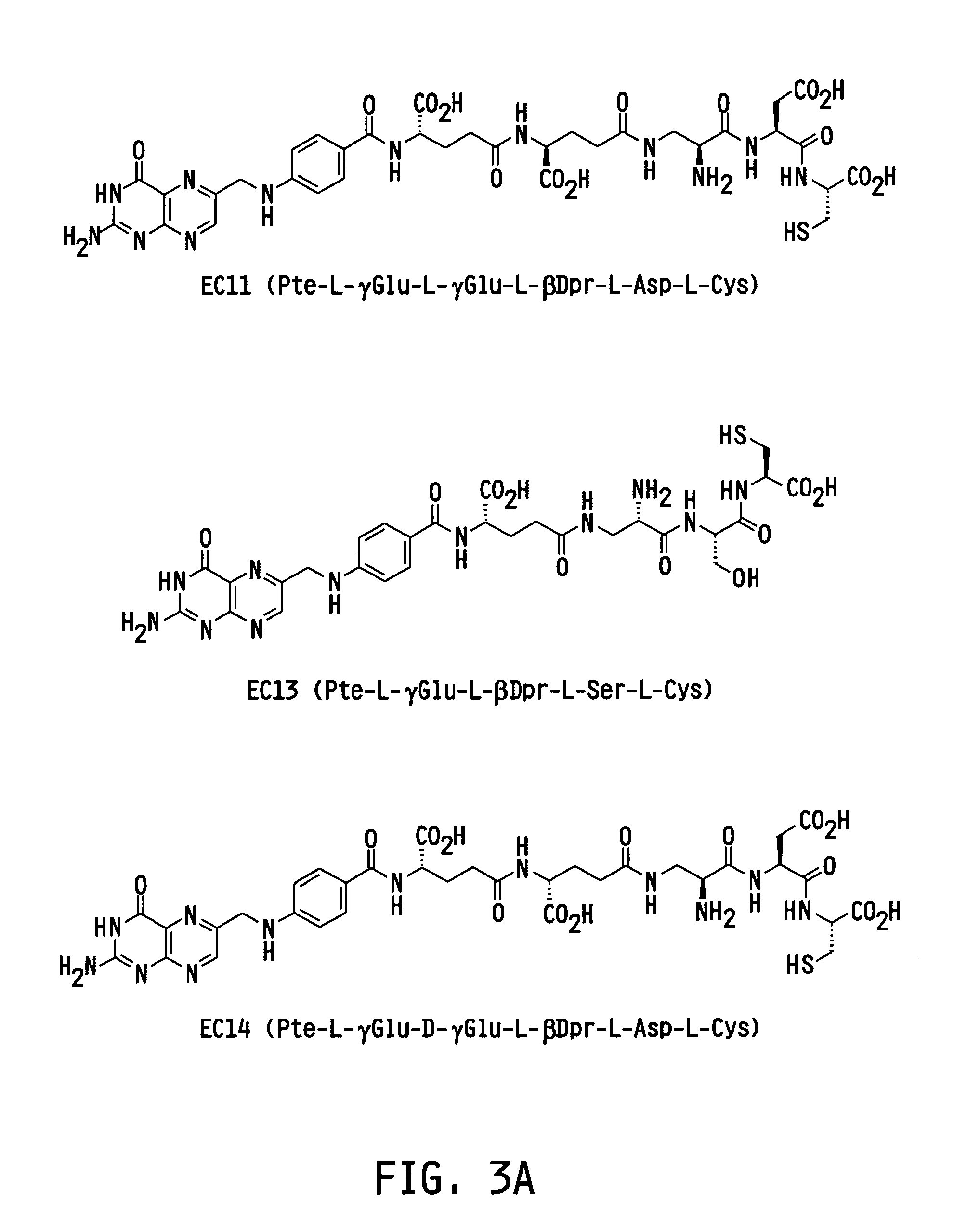
[0077]EC-20 (a folate-linked chelator of 99mTc), was obtained from Endocyte Inc. (West Lafayette, Ind.). EC-20, a low molecular weight (745.2 Da) folate based chelating agent, possesses and rapid radioactive labeling, high binding affinity (Kd˜3 nM) to its target receptor, rapid clearance (I.V. plasma t1 / 2˜4 min), and has minimal side effects. The conjugate consists of a folate targeting ligand tethered to a peptide chelating moiety via a short 2 carbon spacer (FIG. 4). After simple on-site 99mTc labeling, EC-20 imaging can be accomplished in as little as 4 h post injection making this diagnostic agent highly amenable to most clinical settings.

[0078]99mTc-labeled sodium pertechnetate was purchased from Cardinal Health Services (Indianapolis, Ind.). Folic acid and Sephadex G-10 beads were purchased from Sigma / Aldrich (St. Louis, Mo.). Tryptic soy agar (BD 236950) and tryptic soy broth (BD 211825) were purchased from Becton, Dickinson and Company (Franklin Lakes, N.J.). Staph...

example 2

Maintenance of Bacterial Cultures

[0079]Staphylococcus aureus (S. aureus) BAA-934 was propagated according to the protocol supplied by the American Type Culture Collection (ATCC). Briefly, cultures were grown on tryptic soy agar plates or in broth at 37° C. for 24 hours. Tryptic soy culture plates were stored at 2-5° C. for further use. Bacterial enumeratation was based on OD600 measurements. Bacteria were grown to an OD600 of about 1 to about 2 overnight and used directly in tryptic soy broth. Proper Biosafety level 2 guidelines were followed in accordance with CDC and Purdue Biosafety protocols.

example 3

Animal Models of S. Aureus Infection

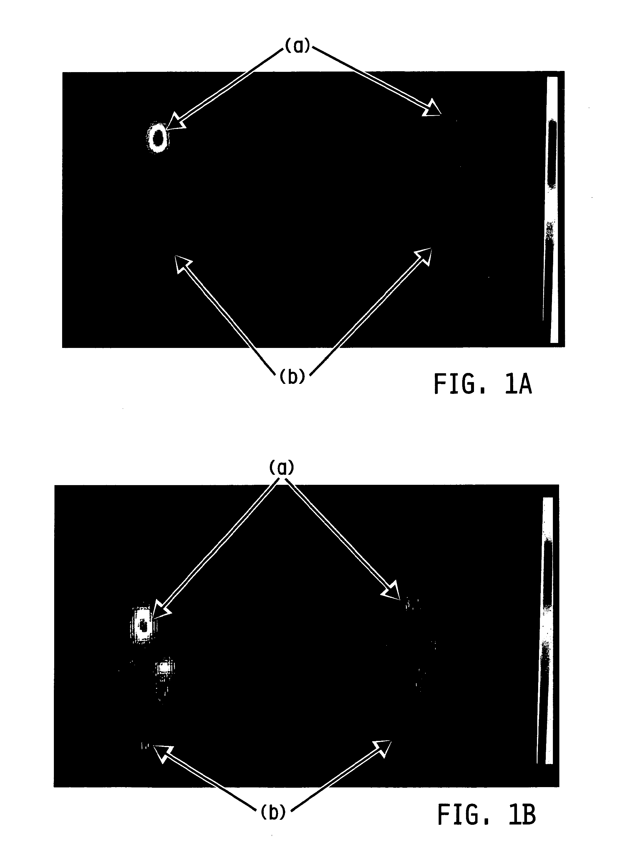
[0080]Infection was induced in either the cuadal muscular (thigh) or flank region in Balb / c (˜20 g) mice as previously described. See Bettegowda, et al. Proc. Natl. Acad. Sci. U.S.A., vol. 102, pp. 1145-1150 (2005) and Bunce, et al. Infect. Immun., vol. 60, pp. 2636-2640 (1992), each incorporated herein by reference. Briefly, on day one, about 2.5×10⁶ CFU to about 1×10⁷ CFU (based on optical density) of S. aureus in 50 μL in tryptic soy broth containing 0.08 g / mL G10 Sephadex beads was injected in the posterior right caudal muscular region (thigh) of the mouse. For induction of the flank model, 107 CFU in 200 μL of tryptic soy agar (containing 0.08 g / mL G10 Sephadex beads) was injected subcutaneously in the right flank. Animals developed abscesses detectable as palpable masses within 24-48 h after inoculation. Animals were given ad libitum access to standard mouse chow and water throughout the course of the study and monitored on a daily basis to ...


PUM

PropertyMeasurementUnit
distanceaaaaaaaaaa
distanceaaaaaaaaaa
distanceaaaaaaaaaa

Abstract

The invention relates to a method for targeting an imaging agent to cells of an animal to detect localized infections. More particularly, localized infections are detected by targeting imaging agents to inflammatory cells having receptors for a vitamin by using vitamin-imaging agent conjugates.

Description

CROSS REFERENCE TO RELATED APPLICATION[0001]This application is a U.S. national counterpart application of international application serial no. PCT / US2008 / 064711 filed May 23, 2008, which claims the benefit of priority under 35 U.S.C. §119(e) of U.S. provisional patent application Ser. No. 60 / 931,753 filed May 25, 2007, the disclosure of which are incorporated herein by reference in their entirety.FIELD OF THE INVENTION[0002]The invention relates to a method for targeting an imaging agent to cells of an animal to detect localized infections. More particularly, localized infections are detected by targeting imaging agents to inflammatory cells having receptors for a ligand, such as by using vitamin-imaging agent conjugates.BACKGROUND AND SUMMARY OF THE INVENTION[0003]Traditional methods for diagnosing infectious diseases rely on microscopic visualization of pathogens in a patient body fluid or tissue sample or the growth of the microorganism in the laboratory with the subsequent iden...

Claims


Application Information

Patent Timeline
Patent Type & Authority Patents(United States)
IPC IPC(8): A61K51/00A61K51/04
CPCA61K51/0459A61P31/00
Inventor LOW, PHILIP STEWARTHENNE, JR., WALTER ANTHONYVARGHESE, BINDUROTHENBUHLER, RYAN
Owner PURDUE RES FOUND INC
Learn More