
Method for differentiating spitz nevi from spitzoid malignant melanoma

a technology of malignant melanoma and spitzoid, applied in the field of protein biology and oncology, can solve the problem of difficult differentiation between benign sn and benign smm

Active Publication Date: 2014-09-02
VANDERBILT UNIV
View PDF2 Cites 3 Cited by
  • Summary
  • Abstract
  • Description
  • Claims
  • Application Information

AI Technical Summary

Benefits of technology

The present invention provides a method for differentiating Spitz nevi from Spitzoid malignant melanoma using mass spectrometry. The method involves comparing the mass spectrometric protein profile of a skin lesion sample from a patient to known profiles of Spitz nevi and Spitzoid malignant melanoma. The method can also involve making a treatment decision for the patient based on the tissue sample. The method may further comprise performing histologic analysis on the skin lesion sample and predicting the patient's survival based on the classification. The invention also provides a kit for performing the method. The technical effect of the invention is to provide a reliable and accurate method for diagnosing and treating melanoma.

Problems solved by technology

However, there are melanocytic lesions, which show conflicting histopathologic criteria and the distinction between a benign SN and SMM may be extremely difficult.
Although gene expression is useful for distinguishing melanocytic nevi from melanomas, it does not always correlate with protein translation and does not account for posttranslational modification (PTM).

Method used


Image

  • Method for differentiating spitz nevi from spitzoid malignant melanoma
  • Method for differentiating spitz nevi from spitzoid malignant melanoma
  • Method for differentiating spitz nevi from spitzoid malignant melanoma

Examples


example 1

Materials and Methods

[0128]Tumor Specimens. The inventors collected archival formalin-fixed, paraffin-embedded (FFPE) tissue samples of SN and SMM from the Yale Spitzoid Neoplasm Repository. The Institutional Review Board at Yale University approved the study. The histopathology of each case was reviewed by the dermatopathologist to confirm the diagnosis. The study included histologically unequivocal SN and primary cutaneous SMM, which were diagnosed initially by a board-certified dermatopathologist. The majority of these cases were seen by multiple dermatopathologists at consensus conference at the time of the initial diagnosis. In addition, all cases chosen for the study underwent confirmatory review by at least 4 other dermatopathologists from Yale Dermatopathology Laboratory. Only cases with an unequivocal consensus diagnosis of SN or melanoma with Spitzoid features (SMM) were included in the study. Histologically ambiguous Spitzoid neoplasms or SN with atypical features were ex...

example 2

Results

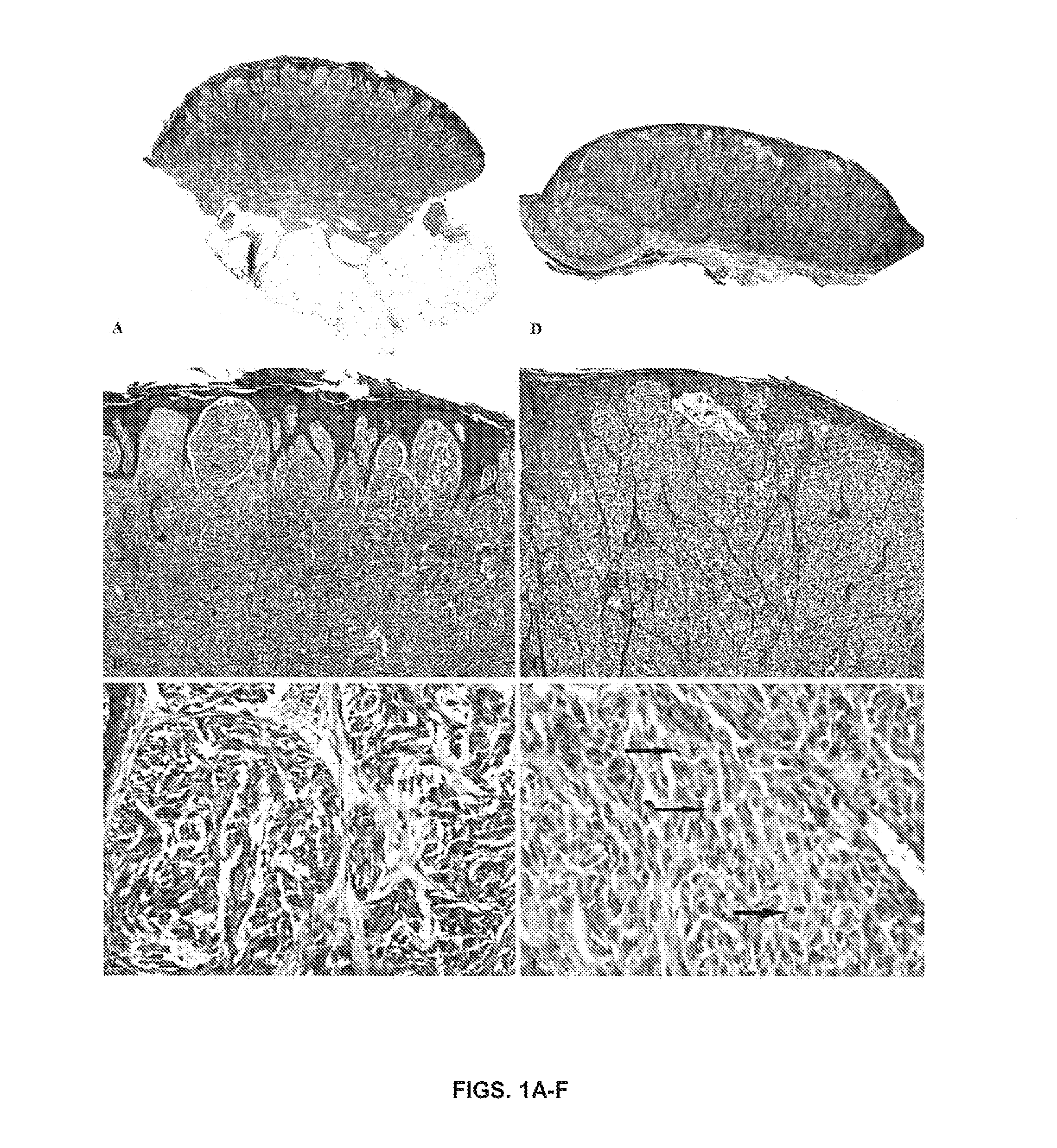
[0135]Characterization of the Study Sample. The cohort of SN came from patients, who ranged from 1 to 48 years of age (mean, 13; SD, 10.2), 30 male and 25 female patients. The lesions were distributed on the head and neck (17), leg (14), back (10), arm (9), buttock (2), abdomen (2), and chest (1). None of the lesions recurred or metastasized, and all patients are alive with a follow-up ranging from 2 to 20 years (mean, 10.7). The SMM cohort comprised patients from 29 to 89 years old (mean, 62; SD, 14), 32 male and 26 female patients. The distribution was as follows: leg (23), back (13), arm (12), scalp (4), chest (2), ear (2), and face (2). The depth of the SMM ranged from 0.75 to 9.0 mm (mean, 3.2 mm). The follow-up ranged from 1 to 21 years (mean, 5). Representative histopathologic features of 1 patient with SN (case #1) and 1 with SMM (case #33) are illustrated in FIGS. 1A-F. A summary of clinical and histopathologic characteristics of patients with SMM is shown in Table 1...


PUM

PropertyMeasurementUnit
diameteraaaaaaaaaa
flow ratesaaaaaaaaaa
diameteraaaaaaaaaa

Abstract

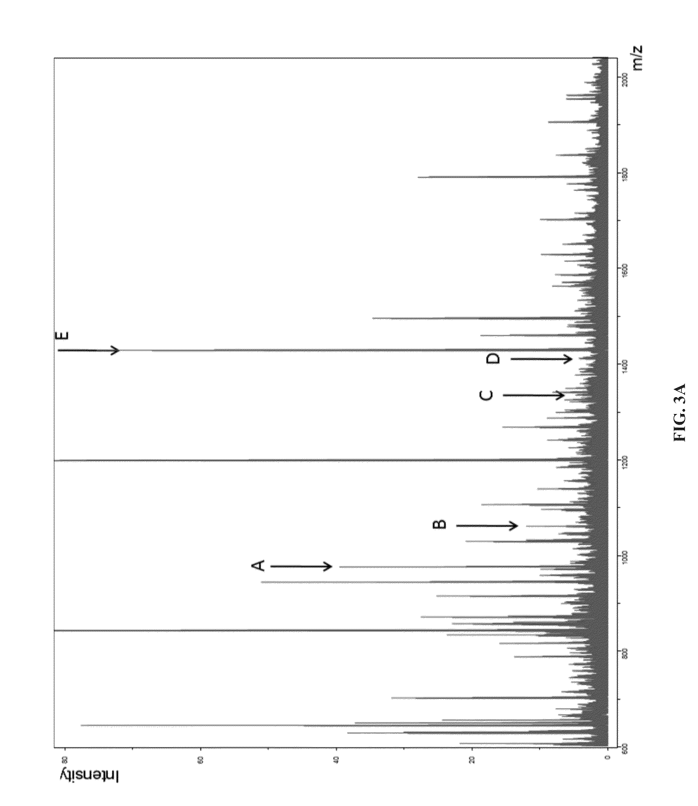
The present invention provides for a mass spectrometry proteomic approach to distinguishing Spitz nevi from Spitzoid malignant melanoma. Histology directed mass spectral profiling allows for targeted analysis of sites of melanocytic lesion within formalin-fixed, paraffin embedded excisional biopsies. The classification system identified 5 peptide peaks, of which two have been identified as originating from vimentin and actin. A sensitivity and specificity for Spitz nevi of 97% and 90%, respectively, were achieved.

Description

[0001]This application claims benefit of priority to U.S. Provisional Application Ser. No. 61 / 593,604, filed Feb. 1, 2012, the entire contents of which are hereby incorporated by reference.[0002]This invention was made with government support under grant no. P50 CA121974 from the National Institutes of Health, and grant no. W81XWH-05-1-0179 from the Army / MRMC. The government has certain rights in the invention.BACKGROUND OF THE INVENTION[0003]1. Field of the Invention[0004]The present invention relates generally to the fields of protein biology and oncology. More particularly, it concerns the classification of skin lesions based on mass spectrometry analysis of proteins from the lesions.[0005]2. Description of Related Art[0006]Ever since the first description of Spitz nevus (SN) by Sophie Spitz in 1948, pathologists and dermatopathologists in particular have been struggling with the distinction between SN and Spitzoid malignant melanoma (SMM). To diagnose a young child with melanoma...

Claims


Application Information

Patent Timeline
Patent Type & Authority Patents(United States)
IPC IPC(8): C12Q1/68
CPCG01N33/5743
Inventor CAPRIOLI, RICHARDSEELEY, ERIN H.
Owner VANDERBILT UNIV
Learn More