
Chimeric phosphorylation indicator

a phosphorylation indicator and chimeric protein technology, applied in the field of chimeric protein phosphorylation indicator, can solve the problems of time-consuming and expensive identification of antibodies that can discriminate between phosphorylated and unphosphorylated forms of proteins, poor temporal and spatial resolution of methods,

Inactive Publication Date: 2014-03-11
RGT UNIV OF CALIFORNIA +1
View PDF7 Cites 1 Cited by
  • Summary
  • Abstract
  • Description
  • Claims
  • Application Information

AI Technical Summary

Benefits of technology

[0029]The sample to be examined for kinase activity can be any sample, including, for example, a sample containing a synthetic product to be examined for kinase or phosphatase activity. In one embodiment, the sample is a biological sample, which can be cell, tissue or organ sample, or an extract of such a sample. In another embodiment, the method is performed on an intact cell, which can be in cell culture or can be in a tissue sample. For such a method, the chimeric phosphorylatable indicator can contain a targeting sequence such as a cell compartmentalization domain that can target the chimeric phosphorylatable indicator to a membrane (e.g., cell membrane or an internal membrane), cytosol, endoplasmic reticulum, mitochondrial matrix, chloroplast lumen, medial trans-Golgi cisternae, a lumen of a lysosome, or a lumen of an endosome. A membrane targeting domain can be a particularly useful to target the chimeric phosphorylation indicators to or near to a cell membrane. A membrane translocating domain can be a particularly useful cell compartmentalization domain is a membrane translocating domain, which can facilitate translocation of the chimeric phosphorylation indicator into an intact cell.

Problems solved by technology

However, the most common currently used technologies such as two dimensional gel electrophoresis, mass spectrometry, co-immunoprecipitation assays, and two-hybrid screens require destroying large numbers of the cells or transferring genes to heterologous organisms.
As such, these methods have poor temporal and spatial resolution, and are insufficient to directly probe physiological functions such as contracture or chemotaxis, which occur on the time scale of milliseconds to minutes.
However, the identification of antibodies that can discriminate between a phosphorylated and unphosphorylated form of a protein is time consuming and expensive.
In addition, the necessary immunocytochemistry is tedious, and is difficult to reassemble into a quantitative time course.
Pertubation of any of these mechanisms disrupts cell function by altering the degree of substrate phosphorylation.
However, GFP-labeling has not proven sufficient to determine the activation state of PKC or PKC-substrate phosphorylation.
This approach has the same advantages and disadvantages as phosphorylation-specific antibodies, including it is applicable to examining endogenous proteins in intact nontransfected tissues, but has poor time resolution and difficulty in generating a continuous time course.
However, although emission from multiple cells may be detectable, the feebleness of bioluminescence would be expected to make the technique difficult or impossible to use with single mammalian cells, especially if high spatial resolution is desired.

Method used


Image

  • Chimeric phosphorylation indicator
  • Chimeric phosphorylation indicator
  • Chimeric phosphorylation indicator

Examples


example 1

Preparation and Characterization of a Chimeric Reporter Protein for a Serine / Threonine Protein Kinase

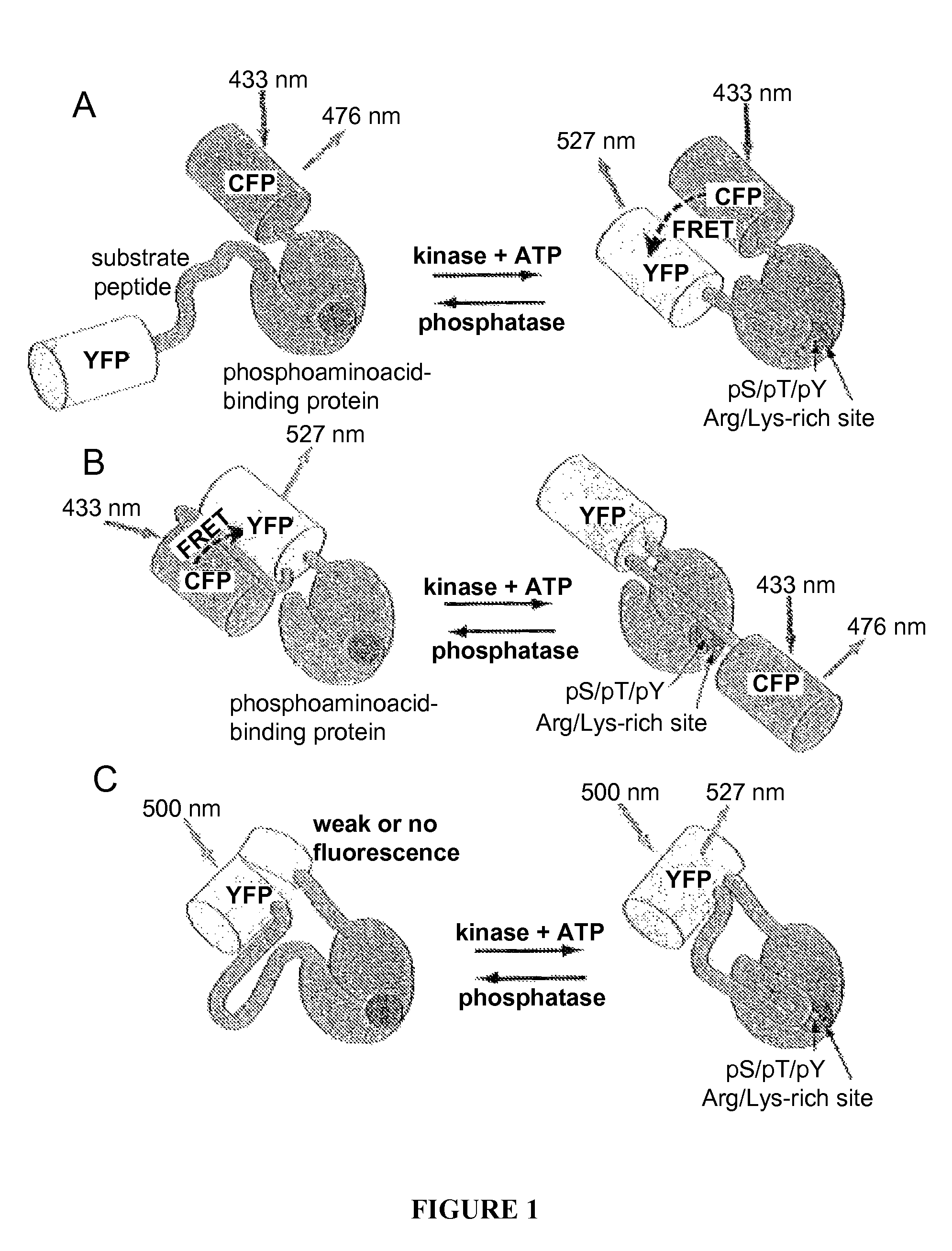
[0165]This example provides a method for preparing a chimeric protein kinase A (PKA; cAMP-dependent protein kinase) reporter protein, and demonstrates that such a chimeric reporter molecule can detect serine / threonine kinase activity.

Plasmid Construction

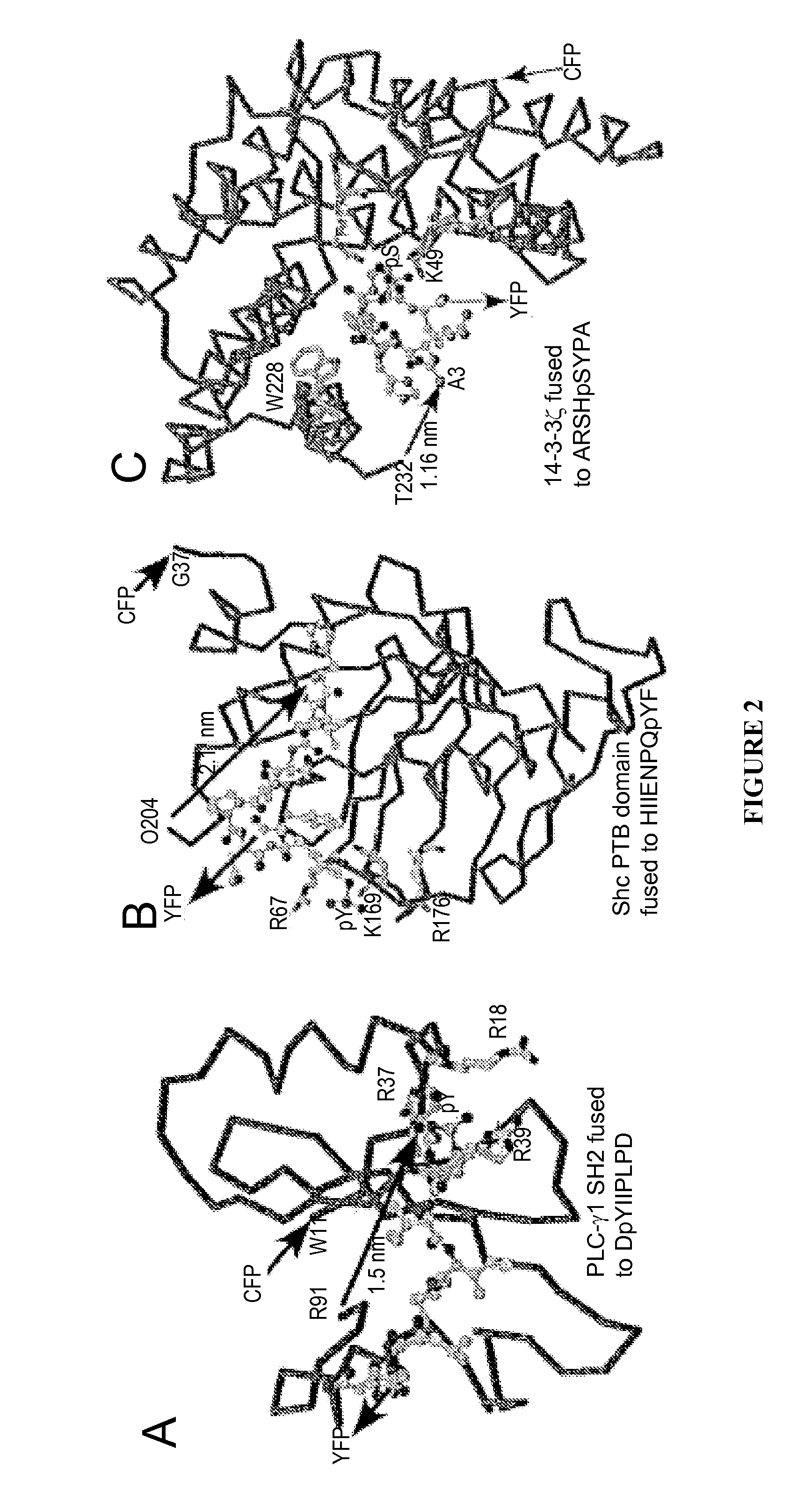
[0166]The PKA chimeric reporter protein was constructed by fusing the enhanced cyan fluorescence protein (1-227; ECFP; SEQ ID NO:6; K26R / F64L / S65T / Y66W / N146I / M153T / V163A / N164H), a truncated version of 14-3-3τ, a modified kemptide and citrine, which is an improved yellow fluorescence protein having a sequence as set forth in SEQ ID NO:10, except containing a Q69M mutation. 14-3-3τ (1-232) was amplified using the cDNA of 14-3-3τ (see GenBank Accession No. D87662) in pcDNA3 vector as the template.

[0167]For PCR, the forward primer had the sequence (SEQ ID NO:26) 5′-GGGCATGCATATGGAGAAGACTGAGCTGATCCAG-3′, and incorporates a Sph I site. The ...

example 2

Preparation and Characterization of Chimeric Reporter Proteins for Detecting Tyrosine Kinase Activity

[0190]This example provides methods for preparing a chimeric src reporter protein and a chimeric EGFR (epidermal growth factor receptor) reporter protein, and demonstrates that such chimeric reporter proteins can detect tyrosine kinase activity.

Preparation of the Chimeric Gene Encoding the Chimeric EGFR Reporter Protein

[0191]The SH2 domain from mouse p52 Shc (see Lanning and Lafuse, Immunogenetics 49:498-504, 1999; GenBank Accession No. AF054823) was amplified by PCR using the following primers:

[0192]

2 EGFR.Fwd-(5′-GCCGCCCGCATGCATTGGTTCCACGGGAAGCTGSEQ ID NO: 38AGCCGG-3′; and)EGFRoptsub.Rev-(5′-TACCATGAGCTCTGATTGCGGAG-CCATGTTCSEQ ID NO: 39ATGTACTCAGCTTCCTCTTCAGGCTTCCCAGATCC-AGAGTGAGACCCCACGGGTTGCTCTAGGCACAG-3′).

[0193]The PCR reaction was assembled as: 67 μl of water, 10 μl of 10×Taq buffer (Promega), 16 μl of 25 mM MgCl2, 2 μl of a 25 mM stock of dNTPs, 1.5 μl of 100 μM EGFR.Fwd prime...

example 3

Identification of Chimeric Phosphorylation Indicators Using High Throughput Screening

[0213]Based on the results described Examples 1 and 2, additional chimeric phosphorylation indicators can be developed and optimized, using a variety of fluorescent proteins, luminescent molecules, kinase or phosphatase substrates, PAABDs, and tetracysteine motifs. High throughput strategies are particularly useful for systematically generating and testing diverse libraries of such constructs (Zhao and Arnold, Curr. Opin. Chem. Biol. 3:284-290, 1997).

[0214]Initial high throughput diversity generation and screening are performed using the exemplified chimeric phosphorylation indicators (Examples 1 and 2), which are active in mammalian cells. Iterative cycles of variegation, for example (see U.S. Pat. No. 5,837,500), can confer additional useful features on these chimeric reporters. Diversity also can be created by a variety of other methods, including, for example, error prone PCR, oligonucleotide-di...


PUM

PropertyMeasurementUnit
Tmaaaaaaaaaa
total volumeaaaaaaaaaa
pHaaaaaaaaaa

Abstract

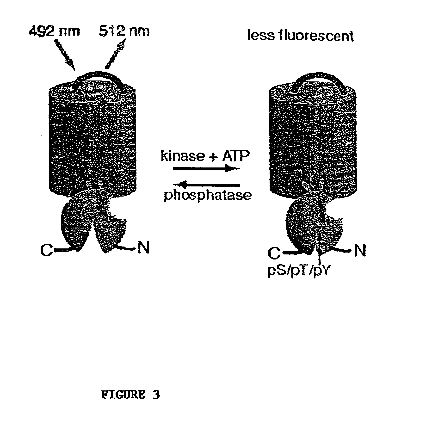
A chimeric phosphorylation indicator (CPI) as provided herein can contain a donor molecule, a phosphorylatable domain, a phosphoaminoacid binding domain (PAABD), and an acceptor molecule. Where the phosphorylatable domain is phosphorylatable by protein kinase C (PKC), the CPI is a c-kinase activity reporter (CKAR). Donor and acceptor molecules may be, independently, fluorescent proteins such as non-oligomerizing fluorescent proteins. A CPI can contain a phosphorylatable polypeptide and a fluorescent protein; the phosphorylatable polypeptide may be contained within the sequence of the fluorescent protein, or the fluorescent protein may be contained within the sequence of the phosphorylatable polypeptide. The spatiotemporal properties of the PKC signal pathway may be tested with CKAR, calcium-sensing fluorophores and FRET-based translocation assays. Polynucleotides encoding such CPIs, and kits containing the indicators and / or the polynucleotides, are provided. A method of using the chimeric phosphorylation indicators to detect a kinase or phosphatase in a sample is provided.

Description

[0001]This application is a continuation-in-part (CIP) of U.S. Ser. No. 09 / 865,291, filed May 24, 2001, now U.S. Pat. No. 6,900,304, which is a CIP of U.S. Ser. No. 09 / 396,003, filed Sep. 13, 1999, which is a continuation (CON) of U.S. Ser. No. 08 / 792,553, filed Jan. 31, 1997 (now U.S. Pat. No. 5,981,200), which is a CIP of U.S. Ser. No. 08 / 594,575, filed Jan. 31, 1996 (now U.S. Pat. No. 6,803,188), the entire contents of each of which is incorporated herein by reference.[0002]This invention was made with government support under Grant No. GM 62114 awarded by the National Institutes of Health. The government has certain rights in this invention.BACKGROUND OF THE INVENTION[0003]1. Field of the Invention[0004]The present invention relates generally to reagents for determining kinase and phosphatase activity, and more specifically to chimeric proteins containing two fluorescent proteins and a phosphorylatable domain, and methods of using such chimeric proteins to detect kinase or phosp...

Claims


Application Information

Patent Timeline
Patent Type & Authority Patents(United States)
IPC IPC(8): G01N33/542C12Q1/48C07K14/47C12P21/04C12Q1/37C07K14/435C12Q1/68C12Q1/42C12N9/12
CPCC07K2319/00C12Q1/485C12Q1/37G01N33/542C07K14/43595G01N2333/43595C07K2319/60C12N9/1205C12Q1/42
Inventor VIOLIN, JONATHAN D.NEWTON, ALEXANDRA C.TSIEN, ROGER Y.ZHANG, JIN
Owner RGT UNIV OF CALIFORNIA
Learn More